1ORR image
Deposition Date 2003-03-14
Release Date 2003-08-26
Last Version Date 2024-02-14
Entry Detail
PDB ID:
1ORR
Keywords:
Title:
Crystal Structure of CDP-Tyvelose 2-Epimerase complexed with NAD and CDP
Biological Source:
Source Organism:
Salmonella typhi (Taxon ID: 601)
Host Organism:
Method Details:
Experimental Method:
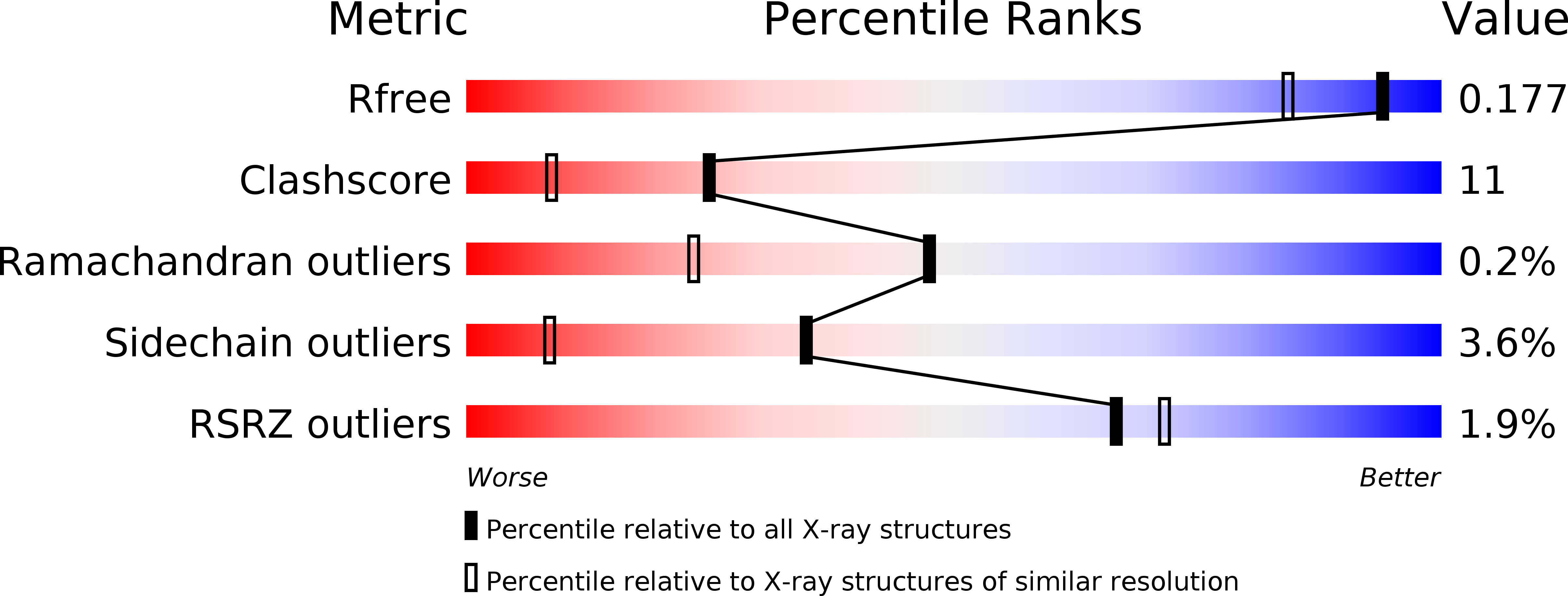
Resolution:
1.50 Å
R-Value Free:
0.22
R-Value Work:
0.17
R-Value Observed:
0.17
Space Group:
P 1 21 1
Macromolecular Entities
Polymer Type:polypeptide(L)
Molecule:CDP-tyvelose-2-epimerase
Gene (Uniprot):rfbE
Chain IDs:A, B, C, D
Chain Length:347
Number of Molecules:4
Biological Source:Salmonella typhi
Primary Citation
High Resolution X-ray Structure of Tyvelose Epimerase from Salmonella typhi
J.Biol.Chem. 278 20874 20881 (2003)
PMID: 12642575 DOI: 10.1074/jbc.M301948200

Abstact

Tyvelose epimerase catalyzes the last step in the biosynthesis of tyvelose by converting CDP-d-paratose to CDP-d-tyvelose. This unusual 3,6-dideoxyhexose occurs in the O-antigens of some types of Gram-negative bacteria. Here we describe the cloning, protein purification, and high-resolution x-ray crystallographic analysis of tyvelose epimerase from Salmonella typhi complexed with CDP. The enzyme from S. typhi is a homotetramer with each subunit containing 339 amino acid residues and a tightly bound NAD+ cofactor. The quaternary structure of the enzyme displays 222 symmetry and can be aptly described as a dimer of dimers. Each subunit folds into two distinct lobes: the N-terminal motif responsible for NAD+ binding and the C-terminal region that harbors the binding site for CDP. The analysis described here demonstrates that tyvelose epimerase belongs to the short-chain dehydrogenase/reductase superfamily of enzymes. Indeed, its active site is reminiscent to that observed for UDP-galactose 4-epimerase, an enzyme that plays a key role in galactose metabolism. Unlike UDP-galactose 4-epimerase where the conversion of configuration occurs about C-4 of the UDP-glucose or UDP-galactose substrates, in the reaction catalyzed by tyvelose epimerase, the inversion of stereochemistry occurs at C-2. On the basis of the observed binding mode for CDP, it is possible to predict the manner in which the substrate, CDP-paratose, and the product, CDP-tyvelose, might be accommodated within the active site of tyvelose epimerase.

Legend

Protein

Chemical

Disease

Primary Citation of related structures