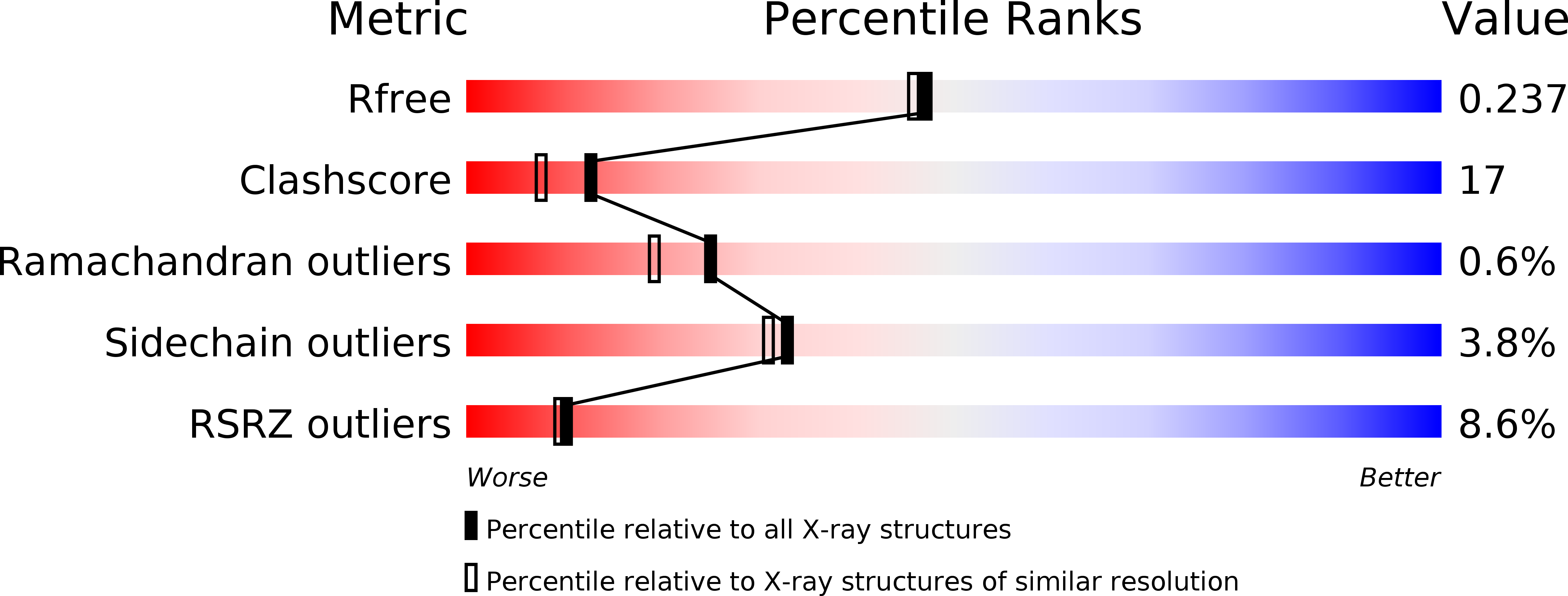
1OR7 image
Deposition Date 2003-03-12
Release Date 2003-04-15
Last Version Date 2024-02-14
Entry Detail
PDB ID:
1OR7
Keywords:
Title:
Crystal Structure of Escherichia coli sigmaE with the Cytoplasmic Domain of its Anti-sigma RseA
Biological Source:
Source Organism:
Escherichia coli (Taxon ID: 562)
Method Details:
Experimental Method:
Resolution:
2.00 Å
R-Value Free:
0.23
R-Value Work:
0.19
R-Value Observed:
0.19
Space Group:
C 1 2 1
Macromolecular Entities
Polymer Type:polypeptide(L)
Molecule:RNA polymerase sigma-E factor
Gene (Uniprot):rpoE
Chain IDs:A, C (auth: B)
Chain Length:194
Number of Molecules:2
Biological Source:Escherichia coli
Polymer Type:polypeptide(L)
Molecule:Sigma-E factor negative regulatory protein
Gene (Uniprot):rseA
Chain IDs:B (auth: C), D (auth: F)
Chain Length:90
Number of Molecules:2
Biological Source:Escherichia coli
Primary Citation
Crystal structure of Escherichia coli sigmaE with the cytoplasmic domain of its anti-sigma RseA.
Mol.Cell 11 1067 1078 (2003)
PMID: 12718891 DOI: 10.1016/S1097-2765(03)00148-5

Abstact

The sigma factors are the key regulators of bacterial transcription. ECF (extracytoplasmic function) sigma's are the largest and most divergent group of sigma(70) family members. ECF sigma's are normally sequestered in an inactive complex by their specific anti-sigma factor, which often spans the inner membrane. Here, we determined the 2 A resolution crystal structure of the Escherichia coli ECF sigma factor sigma(E) in an inhibitory complex with the cytoplasmic domain of its anti-sigma, RseA. Despite extensive sequence variability, the two major domains of sigma(E) are virtually identical in structure to the corresponding domains of other sigma(70) family members. In combination with a model of the sigma(E) holoenzyme and biochemical data, the structure reveals that RseA functions by sterically occluding the two primary binding determinants on sigma(E) for core RNA polymerase.

Legend

Protein

Chemical

Disease

Primary Citation of related structures