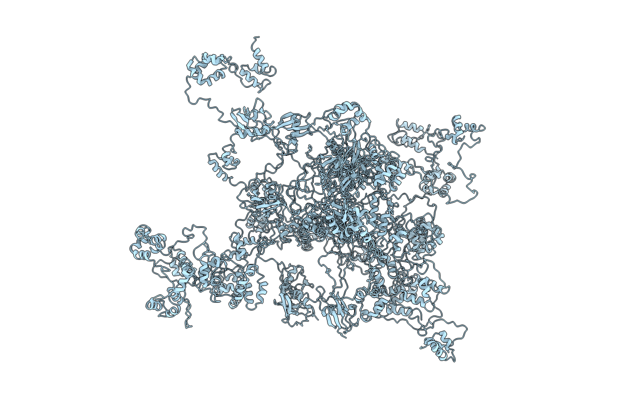
1OQY image
Deposition Date 2003-03-11
Release Date 2003-10-21
Last Version Date 2024-05-22
Entry Detail
PDB ID:
1OQY
Keywords:
Title:
Structure of the DNA repair protein hHR23a
Biological Source:
Source Organism:
Homo sapiens (Taxon ID: 9606)
Method Details:
Experimental Method:
Conformers Calculated:
45
Conformers Submitted:
12
Selection Criteria:
structures with the least restraint violations
Macromolecular Entities
Structures with similar UniProt ID
Protein Blast
Polymer Type:polypeptide(L)
Molecule:UV excision repair protein RAD23 homolog A
Gene (Uniprot):RAD23A
Chain IDs:A
Chain Length:368
Number of Molecules:1
Biological Source:Homo sapiens
Ligand Molecules
Primary Citation
DNA-repair protein hHR23a alters its protein structure upon binding proteasomal subunit S5a
Proc.Natl.Acad.Sci.USA 100 12694 12699 (2003)
PMID: 14557549 DOI: 10.1073/pnas.1634989100

Abstact

The Rad23 family of proteins, including the human homologs hHR23a and hHR23b, stimulates nucleotide excision repair and has been shown to provide a novel link between proteasome-mediated protein degradation and DNA repair. In this work, we illustrate how the proteasomal subunit S5a regulates hHR23a protein structure. By using NMR spectroscopy, we have elucidated the structure and dynamic properties of the 40-kDa hHR23a protein and show it to contain four structured domains connected by flexible linker regions. In addition, we reveal that these domains interact in an intramolecular fashion, and by using residual dipolar coupling data in combination with chemical shift perturbation analysis, we present the hHR23a structure. By itself, hHR23a adopts a closed conformation defined by the interaction of an N-terminal ubiquitin-like domain with two ubiquitin-associated domains. Interestingly, binding of the proteasomal subunit S5a disrupts the hHR23a interdomain interactions and thereby causes it to adopt an opened conformation.

Legend

Protein

Chemical

Disease

Primary Citation of related structures