1OQU image
Deposition Date 2003-03-11
Release Date 2004-04-20
Last Version Date 2024-03-13
Entry Detail
PDB ID:
1OQU
Title:
A protein coordinated tri-nuclear Fe complex formed during soaking of crystals of the ribonucleotide reductase R2F protein from Corynebacterium Ammoniagenes
Biological Source:
Source Organism:
Host Organism:
Method Details:
Experimental Method:
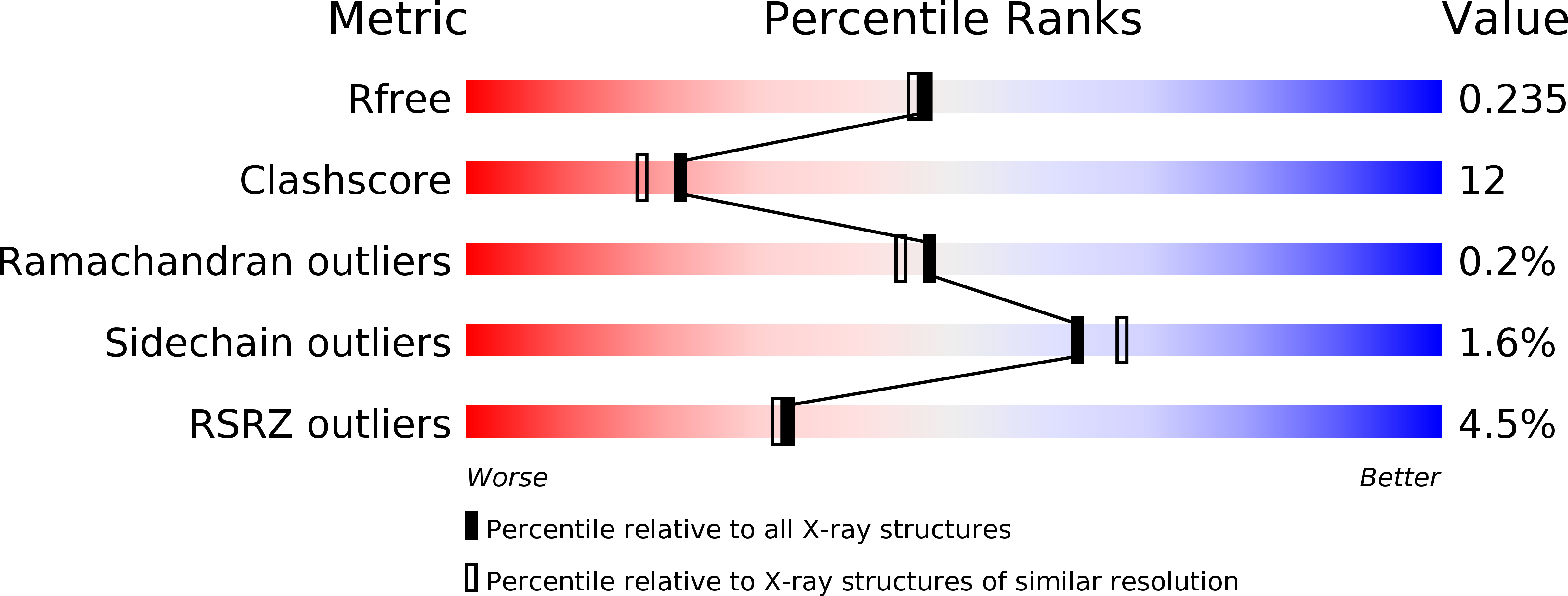
Resolution:
2.00 Å
R-Value Free:
0.23
R-Value Work:
0.18
R-Value Observed:
0.18
Space Group:
P 1 21 1
Macromolecular Entities
Polymer Type:polypeptide(L)
Molecule:ribonucleotide reductase subunit R2F
Gene (Uniprot):nrdF
Chain IDs:A, B, C, D
Chain Length:329
Number of Molecules:4
Biological Source:Corynebacterium ammoniagenes
Primary Citation
A protein carboxylate coordinated oxo-centered tri-nuclear iron complex with possible implications for ferritin mineralization
Febs Lett. 567 179 182 (2004)
PMID: 15178319 DOI: 10.1016/j.febslet.2004.04.068

Abstact

The crystal structure of an oxo-centered tri-nuclear iron complex formed on a protein surface is presented. The cluster forms when crystals of the class Ib ribonucleotide reductase R2 protein from Corynebacterium ammoniagenes are subjected to iron soaking. The tri-iron-oxo complex is coordinated by protein-derived carboxylate ligands arranged in a motif similar to the one found on the inner surface of ferritins and may mimic an early stage in the mineralization of iron in ferritins. In addition, the structure adds to the very limited data on protein-mineral interfaces.

Legend

Protein

Chemical

Disease

Primary Citation of related structures