1OQJ image
Deposition Date 2003-03-10
Release Date 2003-11-11
Last Version Date 2024-02-14
Entry Detail
PDB ID:
1OQJ
Title:
Crystal structure of the SAND domain from glucocorticoid modulatory element binding protein-1 (GMEB1)
Biological Source:
Source Organism:
Homo sapiens (Taxon ID: 9606)
Method Details:
Experimental Method:
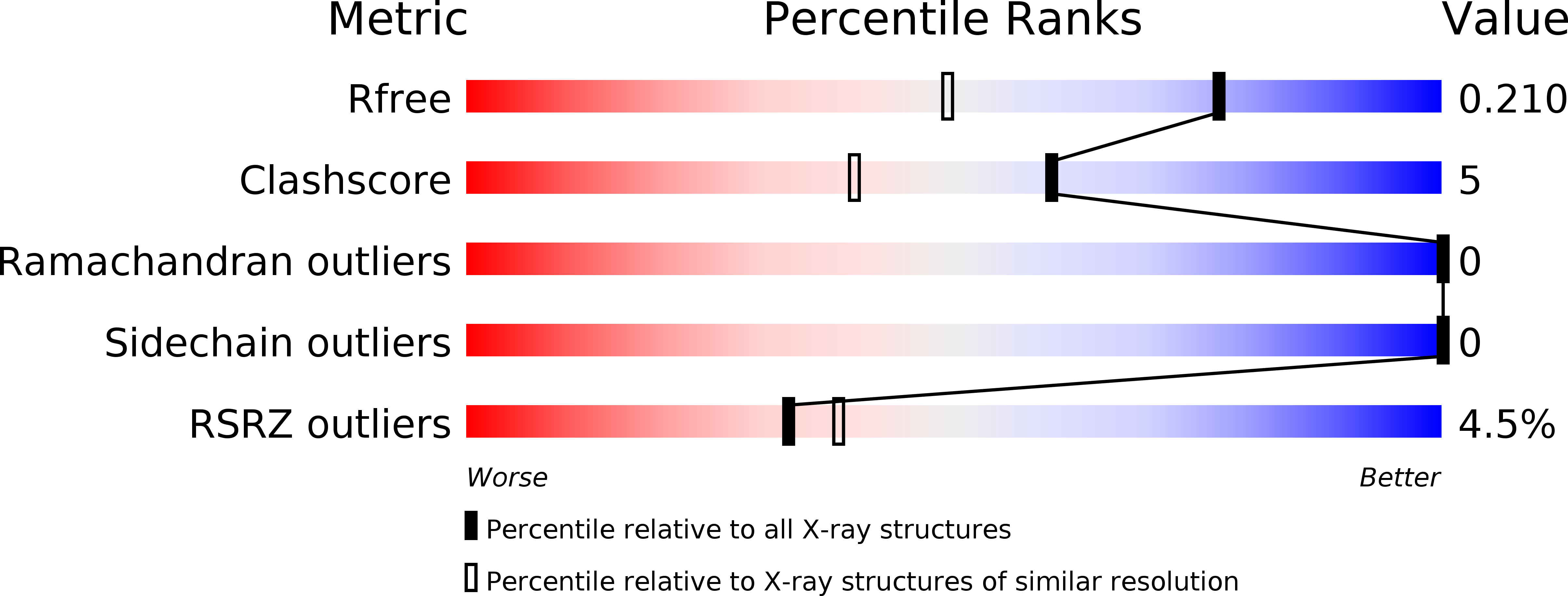
Resolution:
1.55 Å
R-Value Free:
0.21
R-Value Work:
0.19
R-Value Observed:
0.19
Space Group:
P 21 21 21
Macromolecular Entities
Structures with similar UniProt ID
Protein Blast
Polymer Type:polypeptide(L)
Molecule:Glucocorticoid Modulatory Element Binding protein-1
Gene (Uniprot):GMEB1
Chain IDs:A, B
Chain Length:97
Number of Molecules:2
Biological Source:Homo sapiens
Ligand Molecules
Primary Citation
Crystal structure and nuclear magnetic resonance analyses of the SAND domain from glucocorticoid modulatory element binding protein-1 reveals deoxyribonucleic acid and zinc binding regions
MOL.ENDOCRINOL. 17 1283 1295 (2003)
PMID: 12702733 DOI: 10.1210/me.2002-0409

Abstact

The glucocorticoid-modulatory element-binding proteins, GMEB1 and GMEB2, are ubiquitous, multifunctional DNA-binding proteins with important roles in the modulation of transcription upon steroid hormone activation. The GMEB proteins have intrinsic transactivation ability, but also control the glucocorticoid response via direct binding to the glucocorticoid receptor. They are also mandatory host proteins for Parvovirus replication. Here we present the 1.55 A resolution crystal structure of a central portion of GMEB1, encompassing its SAND domain, which shares 80% sequence identity with the GMEB2 SAND domain. We demonstrate that this domain, also present in numerous proteins implicated in chromatin-associated transcriptional regulation, is necessary and sufficient to bind the glucocorticoid-modulatory element (GME) DNA target. We use nuclear magnetic resonance (NMR) and binding studies to map the DNA recognition surface to an alpha-helical region exposing the conserved KDWK motif. Using site-directed mutagenesis, key residues for DNA binding are identified. In contrast to the previously determined NMR structure of the Sp100b SAND domain, we find that the GMEB1 SAND domain also comprises a zinc-binding motif. Although the zinc ion is not necessary for DNA binding, it is found to determine the C-terminal conformation of the GMEB1 SAND domain. We also show that homologous zinc-binding motifs exist in a subset of SAND domain proteins and probe the roles of this novel motif.

Legend

Protein

Chemical

Disease

Primary Citation of related structures