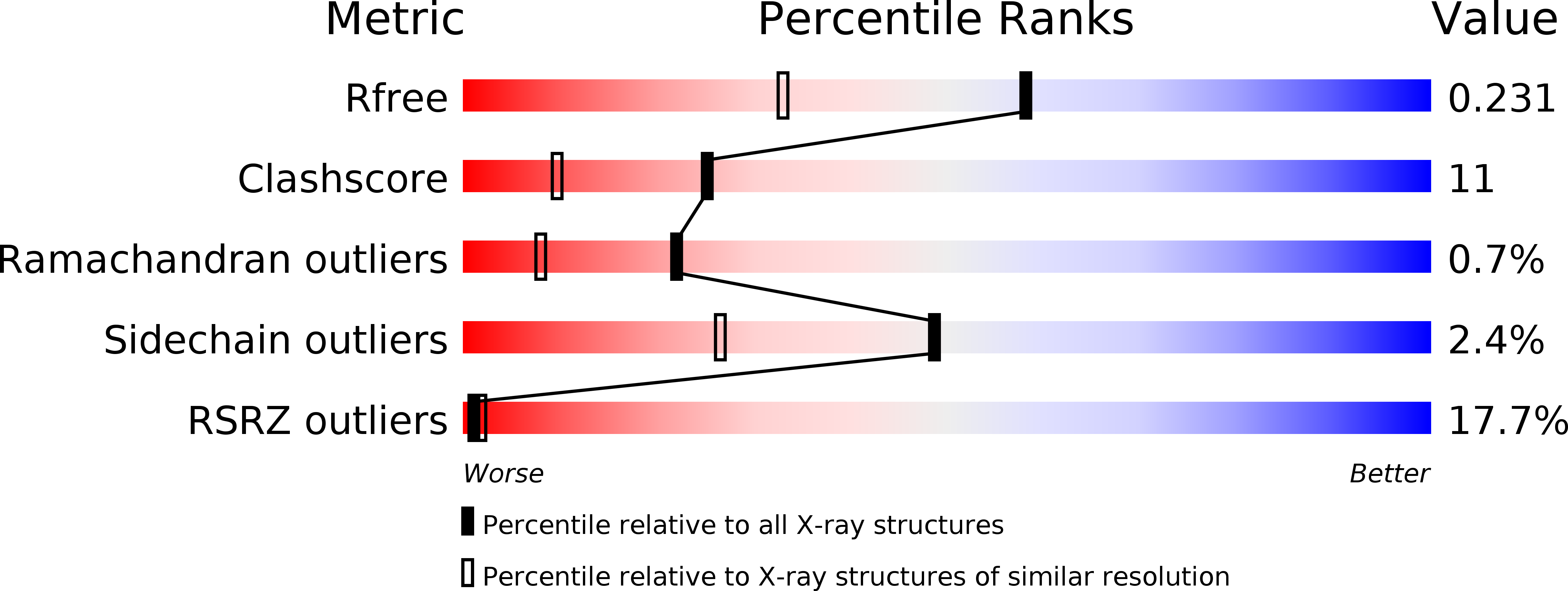
1OPJ image
Deposition Date 2003-03-06
Release Date 2003-04-08
Last Version Date 2024-03-13
Entry Detail
PDB ID:
1OPJ
Keywords:
Title:
Structural basis for the auto-inhibition of c-Abl tyrosine kinase
Biological Source:
Source Organism:
Mus musculus (Taxon ID: 10090)
Host Organism:
Method Details:
Experimental Method:
Resolution:
1.75 Å
R-Value Free:
0.24
R-Value Work:
0.21
R-Value Observed:
0.21
Space Group:
P 1
Macromolecular Entities
Structures with similar UniProt ID
Protein Blast
Polymer Type:polypeptide(L)
Molecule:Proto-oncogene tyrosine-protein kinase ABL1
Gene (Uniprot):Abl1
Chain IDs:A, B
Chain Length:293
Number of Molecules:2
Biological Source:Mus musculus
Primary Citation
Structural basis for the autoinhibition of c-Abl tyrosine kinase
Cell(Cambridge,Mass.) 112 859 871 (2003)
PMID: 12654251 DOI: 10.1016/S0092-8674(03)00194-6

Abstact

c-Abl is normally regulated by an autoinhibitory mechanism, the disruption of which leads to chronic myelogenous leukemia. The details of this mechanism have been elusive because c-Abl lacks a phosphotyrosine residue that triggers the assembly of the autoinhibited form of the closely related Src kinases by internally engaging the SH2 domain. Crystal structures of c-Abl show that the N-terminal myristoyl modification of c-Abl 1b binds to the kinase domain and induces conformational changes that allow the SH2 and SH3 domains to dock onto it. Autoinhibited c-Abl forms an assembly that is strikingly similar to that of inactive Src kinases but with specific differences that explain the differential ability of the drug STI-571/Gleevec/imatinib (STI-571) to inhibit the catalytic activity of Abl, but not that of c-Src.

Legend

Protein

Chemical

Disease

Primary Citation of related structures
Feedback Form
Name
Email
Institute
Feedback