1OP0 image
Deposition Date 2003-03-04
Release Date 2004-05-25
Last Version Date 2024-10-16
Entry Detail
PDB ID:
1OP0
Keywords:
Title:
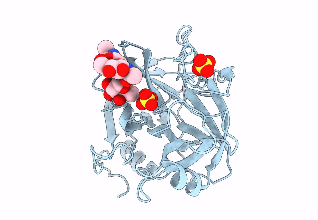
Crystal Structure of AaV-SP-I, a Glycosylated Snake Venom Serine Proteinase from Agkistrodon acutus
Biological Source:
Source Organism:
Method Details:
Experimental Method:
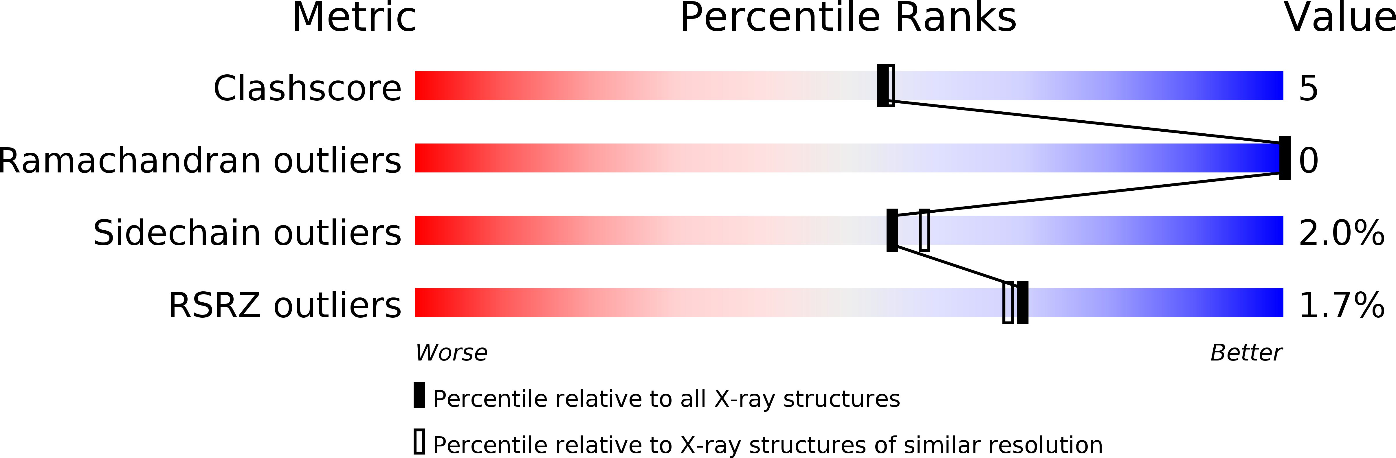
Resolution:
2.00 Å
R-Value Free:
0.20
R-Value Work:
0.18
R-Value Observed:
0.18
Space Group:
P 21 21 21
Macromolecular Entities
Polymer Type:polypeptide(L)
Molecule:Venom serine proteinase
Chain IDs:A
Chain Length:234
Number of Molecules:1
Biological Source:Deinagkistrodon acutus
Primary Citation
Crystal Structures and Amidolytic Activities of Two Glycosylated Snake Venom Serine Proteinases
J.BIOL.CHEM. 280 10524 10529 (2005)
PMID: 15632114 DOI: 10.1074/jbc.M412900200

Abstact

We deduced that Agkistrodon actus venom serine proteinases I and II, previously isolated from the venom of A. acutus (Zhu, Z., Gong, P., Teng, M., and Niu, L. (2003) Acta Crystallogr. Sect. D Biol. Crystallogr. 59, 547-550), are encoded by two almost identical genes, with only the single substitution Asp for Asn at residue 62. Amidolytic assays indicated that they possess slightly different enzymatic properties. Crystal structures of A. actus venom serine proteinases I and II were determined at resolution of 2.0 and 2.1 A with the identification of trisaccharide (NAG(301)-FUC(302)-NAG(303)) and monosaccharide (NAG(301)) residues in them, respectively. The substrate binding sites S3 of the two proteinases appear much shallower than that of Trimeresurus stejnegeri venom plasminogen activator despite the overall structural similarity. Based on structural analysis, we showed that these Asn(35)-linked oligosaccharides collide spatially with some inhibitors, such as soybean trypsin inhibitor, and would therefore hinder their inhibitory binding. Difference of the carbohydrates in both the proteinases might also lead to their altered catalytic activities.

Legend

Protein

Chemical

Disease

Primary Citation of related structures