Deposition Date
2003-03-04
Release Date
2004-04-27
Last Version Date
2023-08-16
Entry Detail
PDB ID:
1OOZ
Keywords:
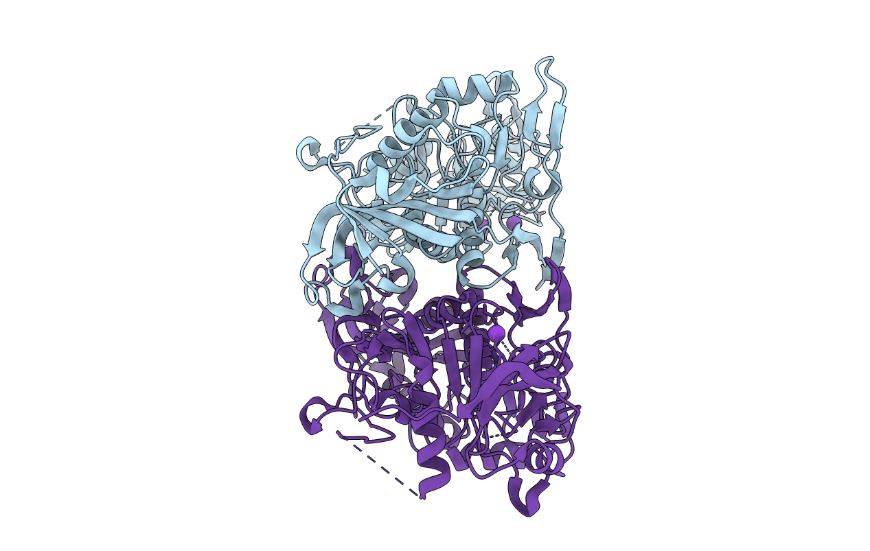
Title:
Deletion mutant of SUCCINYL-COA:3-KETOACID COA TRANSFERASE FROM PIG HEART
Biological Source:
Source Organism(s):
Sus scrofa (Taxon ID: 9823)
Expression System(s):
Method Details:
Experimental Method:
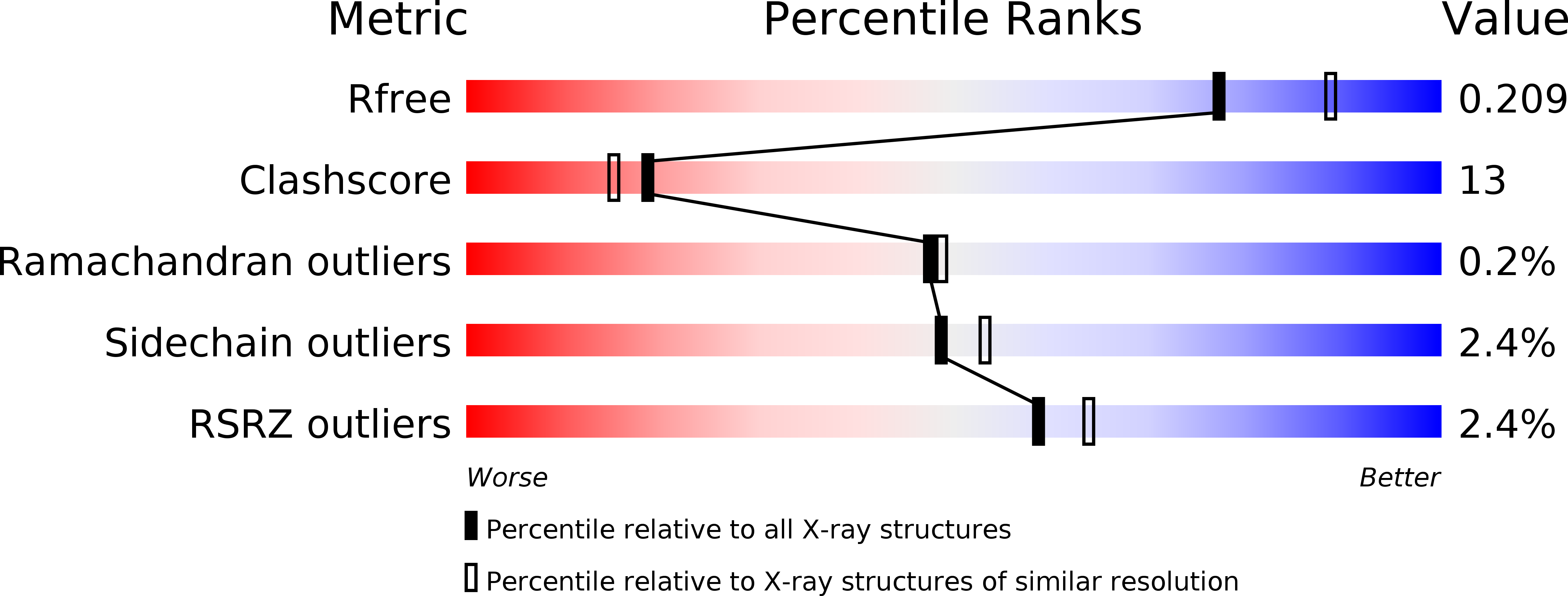
Resolution:
2.10 Å
R-Value Free:
0.21
R-Value Work:
0.16
R-Value Observed:
0.16
Space Group:
P 21 21 2