Deposition Date
2003-02-28
Release Date
2003-08-26
Last Version Date
2023-10-25
Entry Detail
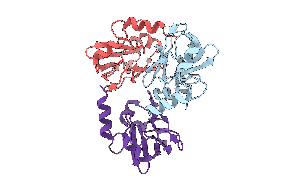
PDB ID:
1ONL
Keywords:
Title:
Crystal structure of Thermus thermophilus HB8 H-protein of the glycine cleavage system
Biological Source:
Source Organism(s):
Thermus thermophilus (Taxon ID: 274)
Expression System(s):
Method Details:
Experimental Method:
Resolution:
2.50 Å
R-Value Free:
0.25
R-Value Work:
0.18
R-Value Observed:
0.18
Space Group:
P 65