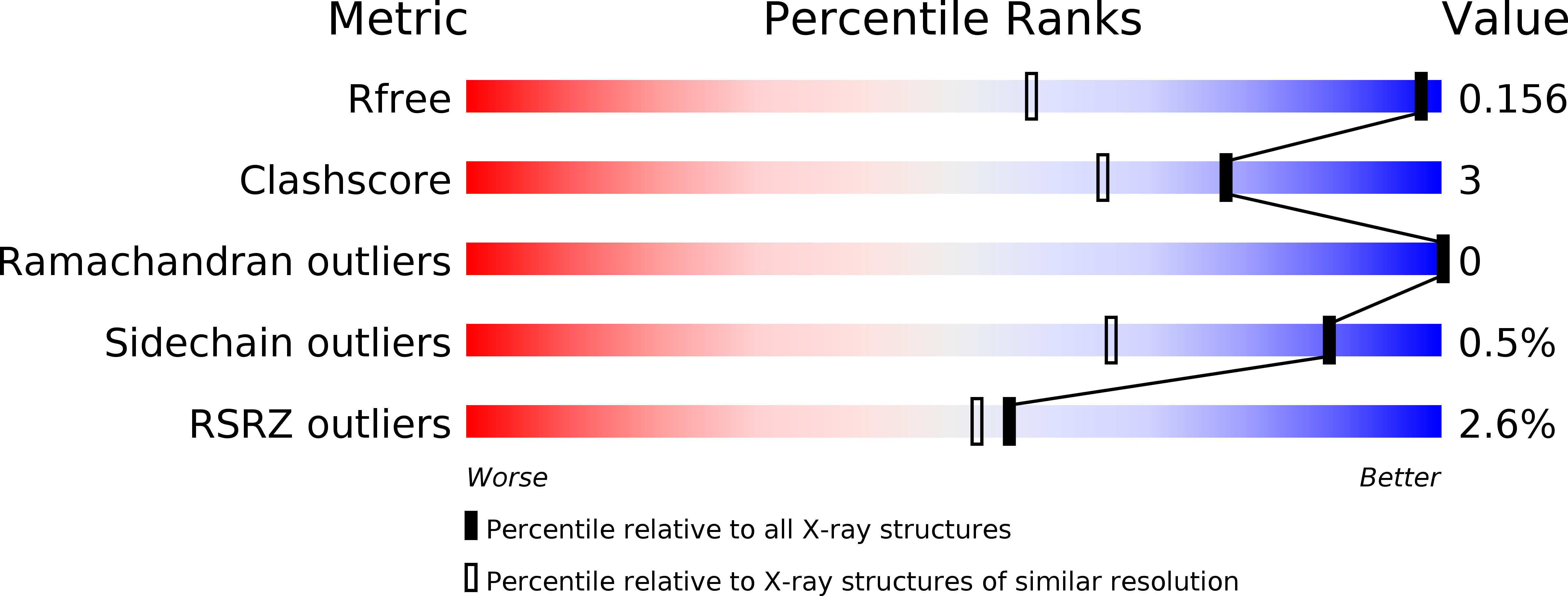
1ONG image
Deposition Date 2003-02-27
Release Date 2003-12-09
Last Version Date 2024-10-30
Entry Detail
PDB ID:
1ONG
Keywords:
Title:
SHV-1 beta-lactamase with a penem inhibitor
Biological Source:
Source Organism:
Method Details:
Experimental Method:
Resolution:
1.10 Å
R-Value Free:
0.18
R-Value Work:
0.14
R-Value Observed:
0.14
Space Group:
P 21 21 21
Macromolecular Entities
Structures with similar UniProt ID
Protein Blast
Polymer Type:polypeptide(L)
Molecule:BETA-LACTAMASE SHV-1
Chain IDs:A
Chain Length:265
Number of Molecules:1
Biological Source:Klebsiella pneumoniae
Primary Citation
Inhibition of Class A and Class C Beta-Lactamases by Penems: Crystallographic Structures of a Novel 1,4-Thiazepine Intermediate
Biochemistry 42 13152 13159 (2003)
PMID: 14609325 DOI: 10.1021/bi034986b

Abstact

A new beta-lactamase inhibitor, a methylidene penem having a 5,6-dihydro-8H-imidazo[2,1-c][1,4]oxazine heterocyclic substituent at the C6 position with a Z configuration, irreversibly inhibits both class A and class C serine beta-lactamases with IC(50) values of 0.4 and 9.0 nM for TEM-1 and SHV-1 (class A), respectively, and 4.8 nM in AmpC (class C) beta-lactamases. The compound also inhibits irreversibly the class C extended-spectrum GC1 beta-lactamase (IC(50) = 6.2 nM). High-resolution crystallographic structures of a reaction intermediate of (5R)-(6Z)-6-(5,6-dihydro-8H-imidazo[2,1-c][1,4]oxazin-2-ylmethylene)-7-oxo-4-thia-1-azabicyclo[3.2.0]hept-2-ene-3-carboxylic acid 1 with the SHV-1 beta-lactamase and with the GC1 beta-lactamase have been determined by X-ray diffraction to resolutions of 1.10 and 1.38 A, respectively. The two complexes were refined to crystallographic R-factors (R(free)) of 0.141 (0.186) and 0.138 (0.202), respectively. Cryoquenching of the reaction of 1 with each beta-lactamase crystal produced a common, covalently bound intermediate. After acylation of the serine, a nucleophilic attack by the departing thiolate on the C6' atom yielded a novel seven-membered 1,4-thiazepine ring having R stereochemistry at the new C7 moiety. The orientation of this ring in each complex differs by a 180 degrees rotation about the bond to the acylated serine. The acyl ester bond is stabilized to hydrolysis through resonance stabilization with the dihydrothiazepine ring and by low occupancy or disorder of hydrolytic water molecules. In the class A complex, the buried water molecule on the alpha-face of the ester bond appears to be loosely bound or absent. In the class C complex, a water molecule on the beta-face is disordered and poorly activated for hydrolysis. Here, the acyl intermediate is unable to assist its own hydrolysis, as is thought to occur with many class C substrates.

Legend

Protein

Chemical

Disease

Primary Citation of related structures