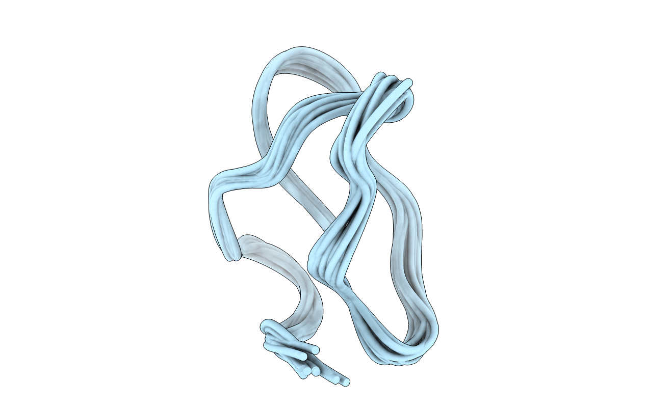
The three-dimensional solution structure of omega-conotoxin MVIIA, a 25-mer peptide antagonist of N-type calcium channels, was determined by two-dimensional 1H NMR spectroscopy with simulated annealing calculations. A total of 13 converged structures of omega-conotoxin MVIIA were obtained on the basis of 273 experimental constraints, including 232 distance constraints obtained from nuclear Overhauser effect (NOE) connectivities, 22 torsion angle (phi, chi 1) constraints, and 19 constraints associated with hydrogen bonds and disulfide bonds. The atomic root mean square difference about the averaged coordinate positions is 0.47 +/- 0.08 A for the backbone atoms (N, C alpha, C) and 1.27 +/- 0.14 A for all heavy atoms of the entire peptide. The molecular structure of omega-conotoxin MVIIA is composed of a short triple-stranded antiparallel beta-sheet. The overall beta-sheet topology is +2x, -1, which is the same as that reported for omega-conotoxin GVIA, another N-type calcium channel blocker. The orientation of beta-stranded structure is similar to each other, suggesting that the conserved disulfide bond combination is essential for the molecular folding. We have recently determined by using alanine substitution analyses that Tyr 13 is essential for the activity of both toxins. On the basis of functional and structural analysis, it is shown that both omega-conotoxin MVIIA and GVIA retain a similar conformation to locate Tyr 13 in the appropriate position to allow binding to N-type calcium channels. These results provide a molecular basis for understanding the mechanism of calcium channel modulation through the toxin-channel interaction and insight into the discrimination of different subtypes of calcium channels.
Legend
Protein
Chemical
Disease