Deposition Date
1998-02-09
Release Date
1998-05-27
Last Version Date
2024-05-22
Entry Detail
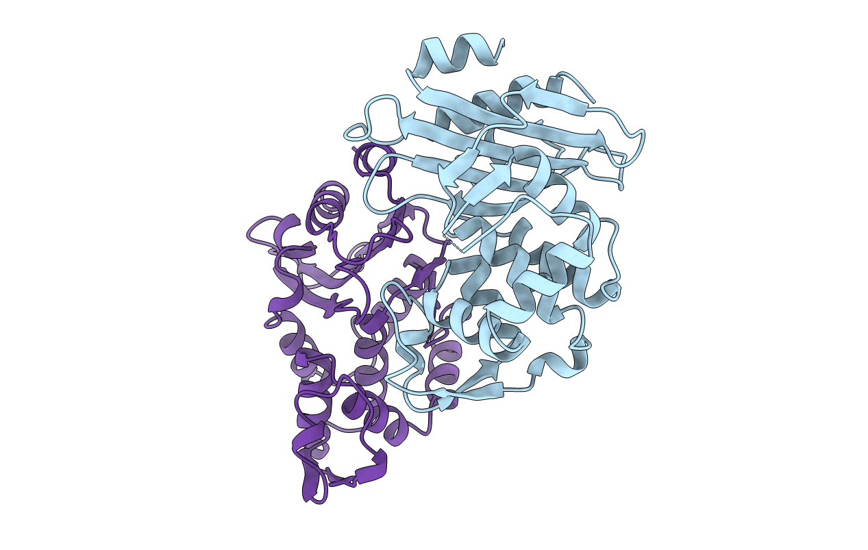
PDB ID:
1OME
Keywords:
Title:
CRYSTAL STRUCTURE OF THE OMEGA LOOP DELETION MUTANT (RESIDUES 163-178 DELETED) OF BETA-LACTAMASE FROM STAPHYLOCOCCUS AUREUS PC1
Biological Source:
Source Organism(s):
Staphylococcus aureus (Taxon ID: 1280)
Expression System(s):
Method Details:
Experimental Method:
Resolution:
2.30 Å
R-Value Free:
0.29
R-Value Work:
0.19
R-Value Observed:
0.19
Space Group:
P 1 21 1