Deposition Date
1990-04-19
Release Date
1991-07-15
Last Version Date
2024-02-14
Entry Detail
PDB ID:
1OMD
Keywords:
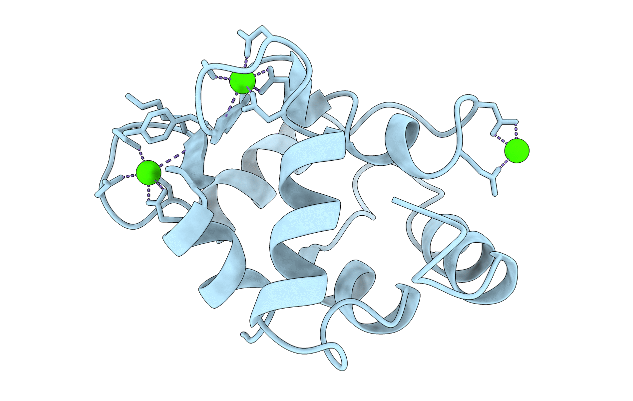
Title:
STRUCTURE OF ONCOMODULIN REFINED AT 1.85 ANGSTROMS RESOLUTION. AN EXAMPLE OF EXTENSIVE MOLECULAR AGGREGATION VIA CA2+
Biological Source:
Source Organism(s):
Rattus norvegicus (Taxon ID: 10116)
Method Details:
Experimental Method:
Resolution:
1.85 Å
R-Value Observed:
0.16
Space Group:
P 21 21 21