1OMC image
Deposition Date 1993-04-28
Release Date 1994-01-31
Last Version Date 2025-03-26
Entry Detail
PDB ID:
1OMC
Title:
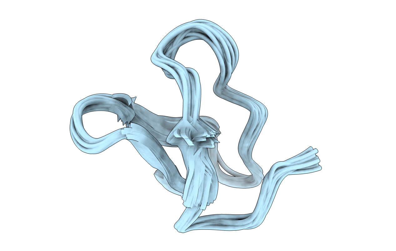
SOLUTION STRUCTURE OF OMEGA-CONOTOXIN GVIA USING 2-D NMR SPECTROSCOPY AND RELAXATION MATRIX ANALYSIS
Biological Source:
Source Organism:
Method Details:
Experimental Method:
Conformers Submitted:
21
Macromolecular Entities
Polymer Type:polypeptide(L)
Molecule:OMEGA-CONOTOXIN GVIA
Chain IDs:A
Chain Length:28
Number of Molecules:1
Biological Source:Conus geographus
Modified Residue
Compound ID Chain ID Parent Comp ID Details 2D Image
HYP A PRO 4-HYDROXYPROLINE
Ligand Molecules
Primary Citation
Solution structure of omega-conotoxin GVIA using 2-D NMR spectroscopy and relaxation matrix analysis.
Biochemistry 32 7396 7405 (1993)
PMID: 8338837 DOI: 10.1021/bi00080a009

Abstact

We report here the solution structure of omega-conotoxin GVIA, a peptide antagonist of the N-type neuronal voltage-sensitive calcium channel. The structure was determined using two-dimensional NMR in combination with distance geometry and restrained molecular dynamics. The full relaxation matrix analysis program MARDIGRAS was used to generate maximum and minimum distance restraints from the crosspeak intensities in NOESY spectra. The 187 restraints obtained were used in conjunction with 23 angle restraints from vicinal coupling constants as input for the structure calculations. The backbones of the best 21 structures match with an average pairwise RMSD of 0.58 A. The structures contain a short segment of triple-stranded beta-sheet involving residues 6-8, 18-21, and 24-27, making this the smallest published peptide structure to contain a triple-stranded beta-sheet. Conotoxins have been shown to be effective neuroprotective agents in animal models of brain ischemia. Our results should aid in the design of novel nonpeptide compounds with potential therapeutic utility.

Legend

Protein

Chemical

Disease

Primary Citation of related structures