Deposition Date
2003-08-19
Release Date
2003-09-11
Last Version Date
2024-11-13
Entry Detail
PDB ID:
1OLZ
Keywords:
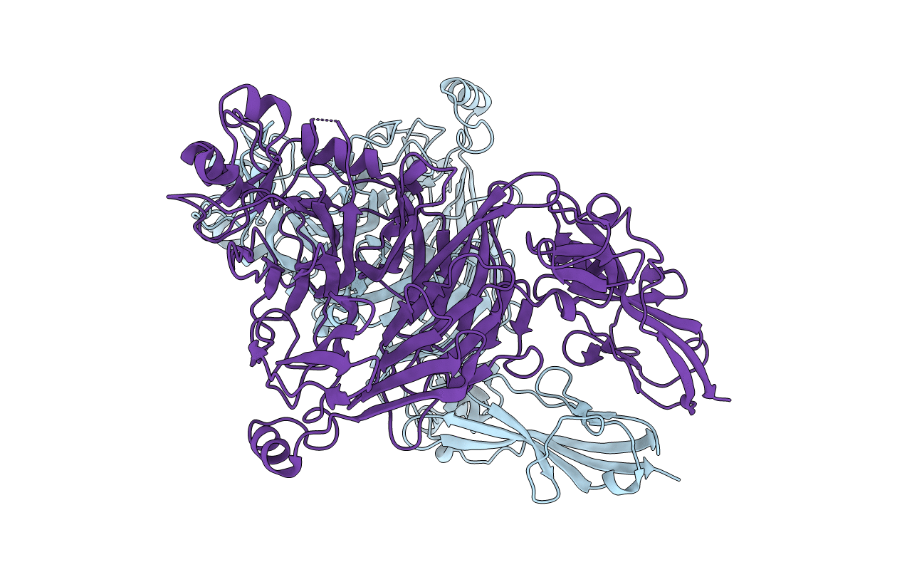
Title:
The ligand-binding face of the semaphorins revealed by the high resolution crystal structure of SEMA4D
Biological Source:
Source Organism(s):
Homo sapiens (Taxon ID: 9606)
Expression System(s):
Method Details:
Experimental Method:
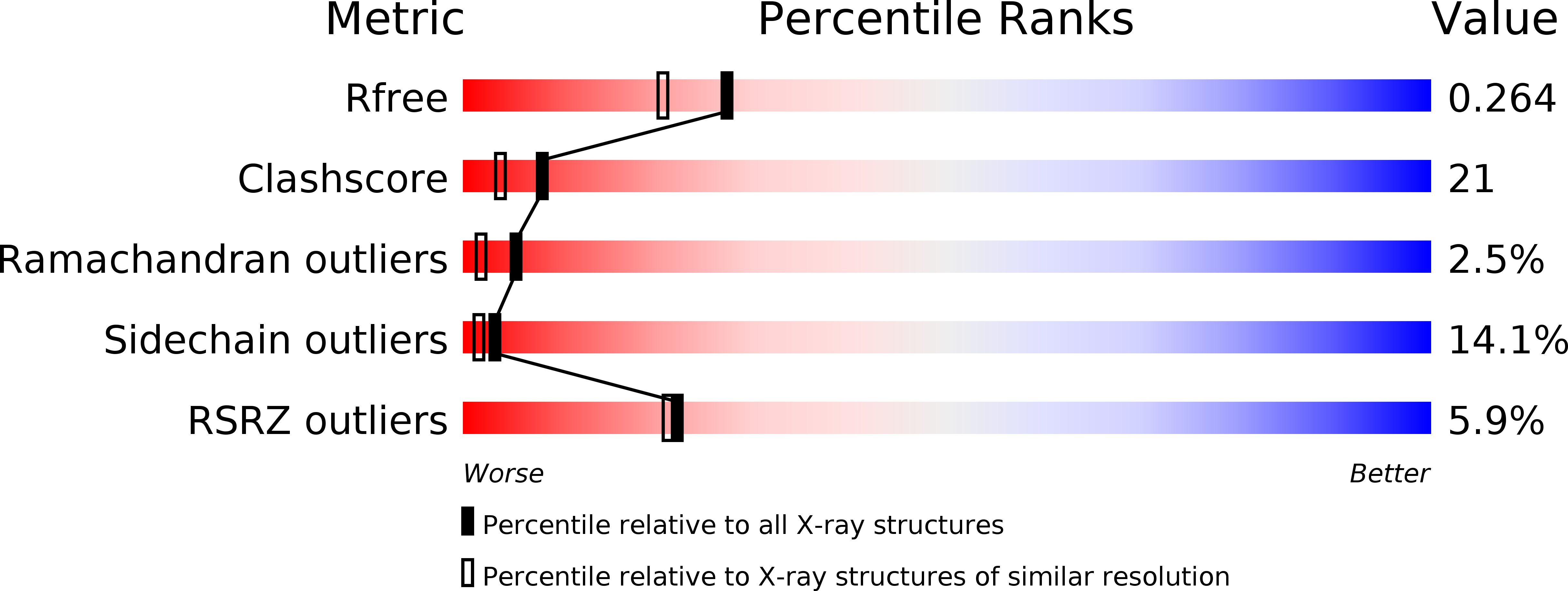
Resolution:
2.00 Å
R-Value Free:
0.27
R-Value Work:
0.20
R-Value Observed:
0.20
Space Group:
P 1