1OL7 image
Deposition Date 2003-08-06
Release Date 2003-10-30
Last Version Date 2024-11-20
Entry Detail
PDB ID:
1OL7
Keywords:
Title:
Structure of Human Aurora-A 122-403 phosphorylated on Thr287, Thr288
Biological Source:
Source Organism:
HOMO SAPIENS (Taxon ID: 9606)
Host Organism:
Method Details:
Experimental Method:
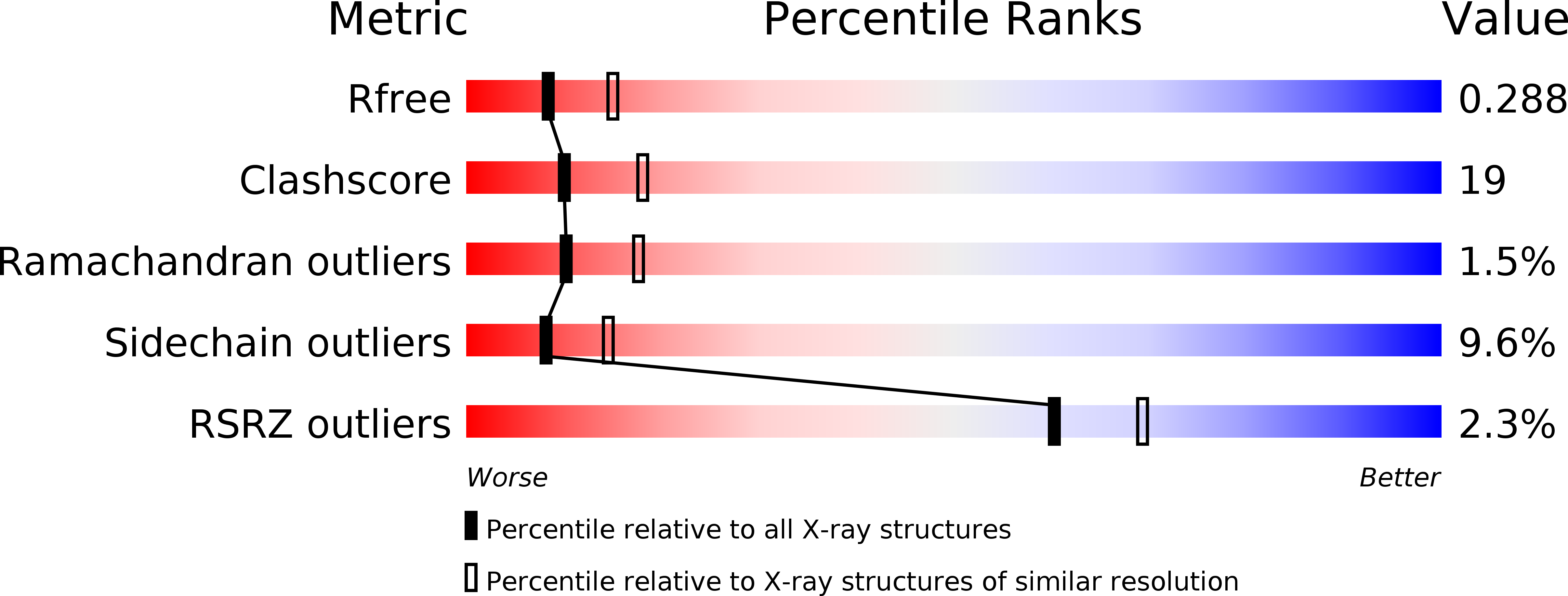
Resolution:
2.75 Å
R-Value Free:
0.29
R-Value Work:
0.25
R-Value Observed:
0.25
Space Group:
P 61 2 2
Macromolecular Entities
Polymer Type:polypeptide(L)
Molecule:SERINE/THREONINE KINASE 6
Gene (Uniprot):AURKA
Chain IDs:A
Chain Length:282
Number of Molecules:1
Biological Source:HOMO SAPIENS
Modified Residue
Compound ID Chain ID Parent Comp ID Details 2D Image
TPO A THR PHOSPHOTHREONINE
Primary Citation
Structural Basis of Aurora-A Activation by Tpx2 at the Mitotic Spindle
Mol.Cell 12 851 ? (2003)
PMID: 14580337 DOI: 10.1016/S1097-2765(03)00392-7

Abstact

Aurora-A is an oncogenic kinase essential for mitotic spindle assembly. It is activated by phosphorylation and by the microtubule-associated protein TPX2, which also localizes the kinase to spindle microtubules. We have uncovered the molecular mechanism of Aurora-A activation by determining crystal structures of its phosphorylated form both with and without a 43 residue long domain of TPX2 that we identified as fully functional for kinase activation and protection from dephosphorylation. In the absence of TPX2, the Aurora-A activation segment is in an inactive conformation, with the crucial phosphothreonine exposed and accessible for deactivation. Binding of TPX2 triggers no global conformational changes in the kinase but pulls on the activation segment, swinging the phosphothreonine into a buried position and locking the active conformation. The recognition between Aurora-A and TPX2 resembles that between the cAPK catalytic core and its flanking regions, suggesting this molecular mechanism may be a recurring theme in kinase regulation.

Legend

Protein

Chemical

Disease

Primary Citation of related structures