1OL2 image
Deposition Date 2003-08-05
Release Date 2003-12-11
Last Version Date 2023-12-13
Entry Detail
PDB ID:
1OL2
Title:
Cyclin A binding groove inhibitor H-Arg-Arg-Leu-Asn-(p-F-Phe)-NH2
Biological Source:
Source Organism:
Host Organism:
Method Details:
Experimental Method:
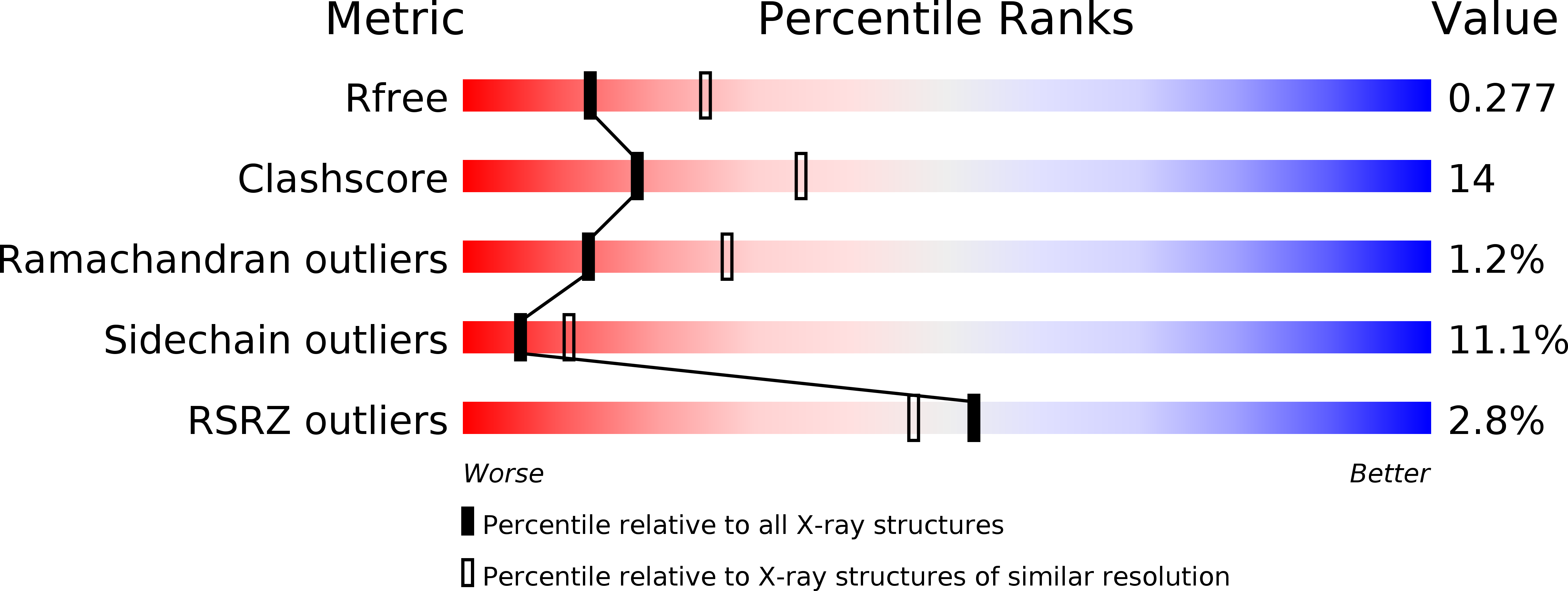
Resolution:
2.60 Å
R-Value Free:
0.29
R-Value Work:
0.19
R-Value Observed:
0.19
Space Group:
P 21 21 21
Macromolecular Entities
Polymer Type:polypeptide(L)
Molecule:CELL DIVISION PROTEIN KINASE 2
Gene (Uniprot):CDK2
Chain IDs:A, C
Chain Length:298
Number of Molecules:2
Biological Source:HOMO SAPIENS
Polymer Type:polypeptide(L)
Molecule:CYCLIN A2
Gene (Uniprot):CCNA2
Chain IDs:B, D
Chain Length:260
Number of Molecules:2
Biological Source:HOMO SAPIENS
Polymer Type:polypeptide(L)
Molecule:ARG-ARG-LEU-ASN-PFF-NH2
Chain IDs:E, F
Chain Length:6
Number of Molecules:2
Biological Source:SYNTHETIC CONSTRUCT
Primary Citation
Insights Into Cyclin Groove Recognition. Complex Crystal Structures and Inhibitor Design Through Ligand Exchange
Structure 11 1537 ? (2003)
PMID: 14656438 DOI: 10.1016/J.STR.2003.11.006

Abstact

Inhibition of CDK2/CA (cyclin-dependent kinase 2/cyclin A complex) activity through blocking of the substrate recognition site in the cyclin A subunit has been demonstrated to be an effective method for inducing apoptosis in tumor cells. We have used the cyclin binding motif (CBM) present in the tumor suppressor proteins p21(WAF1) and p27(KIP1) as a template to optimize the minimal sequence necessary for CDK2/CA inhibition. A series of peptides were prepared, containing nonnatural amino acids, which possess nano- to micromolar CDK2-inhibitory activity. Here we present X-ray structures of the protein complex CDK2/CA, together with the cyclin groove-bound peptides H-Ala-Ala-Abu-Arg-Ser-Leu-Ile-(p-F-Phe)-NH(2) (peptide 1), H-Arg-Arg-Leu-Ile-Phe-NH(2) (peptide 2), Ac-Arg-Arg-Leu-Asn-(m-Cl-Phe)-NH(2) (peptide 3), H-Arg-Arg-Leu-Asn-(p-F-Phe)-NH(2) (peptide 4), and H-Cit-Cit-Leu-Ile-(p-F-Phe)-NH(2) (peptide 5). Some of the peptide complexes presented here were obtained through the novel technique of ligand exchange within protein crystals. This method may find general application for obtaining complex structures of proteins with surface-bound ligands.

Legend

Protein

Chemical

Disease