Deposition Date
2003-07-31
Release Date
2003-10-24
Last Version Date
2023-12-13
Entry Detail
PDB ID:
1OKX
Keywords:
Title:
Binding Structure of Elastase Inhibitor Scyptolin A
Biological Source:
Source Organism(s):
SUS SCROFA (Taxon ID: 9823)
SCYTONEMA HOFMANNI (Taxon ID: 34078)
SCYTONEMA HOFMANNI (Taxon ID: 34078)
Method Details:
Experimental Method:
Resolution:
2.80 Å
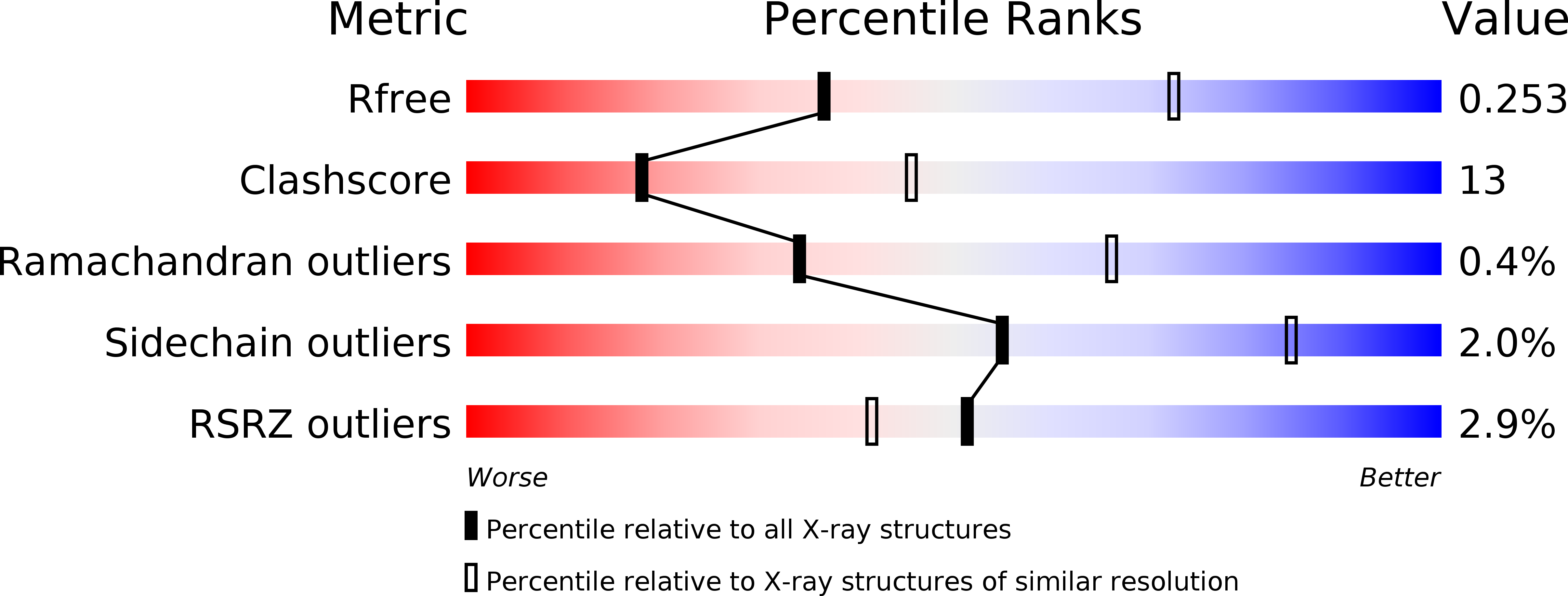
R-Value Free:
0.26
R-Value Work:
0.21
R-Value Observed:
0.21
Space Group:
P 6 2 2