Deposition Date
2003-07-28
Release Date
2003-09-11
Last Version Date
2024-11-13
Entry Detail
PDB ID:
1OKQ
Keywords:
Title:
LAMININ ALPHA 2 CHAIN LG4-5 DOMAIN PAIR, CA1 SITE MUTANT
Biological Source:
Source Organism(s):
MUS MUSCULUS (Taxon ID: 10090)
Expression System(s):
Method Details:
Experimental Method:
Resolution:
2.80 Å
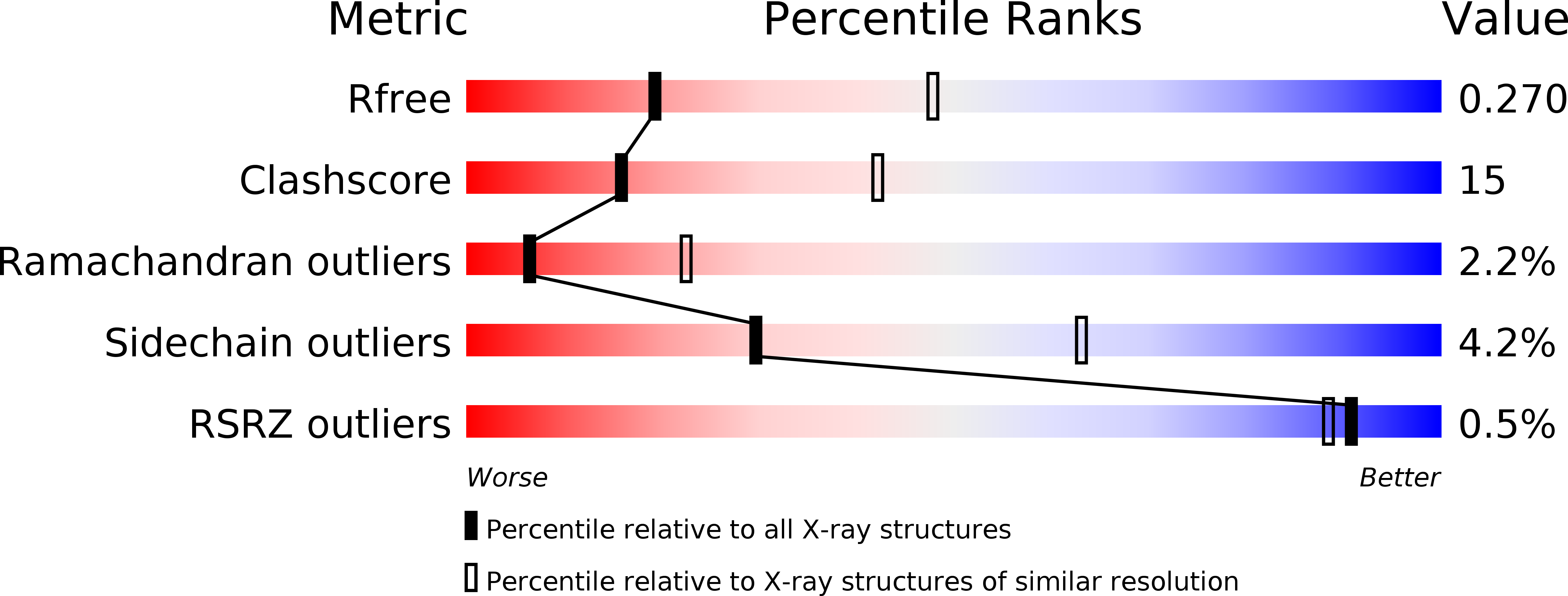
R-Value Free:
0.27
R-Value Work:
0.22
R-Value Observed:
0.22
Space Group:
C 2 2 21