1OKD image
Deposition Date 2003-07-22
Release Date 2003-08-28
Last Version Date 2024-10-16
Entry Detail
PDB ID:
1OKD
Title:
NMR-structure of tryparedoxin 1
Biological Source:
Source Organism:
Host Organism:
Method Details:
Experimental Method:
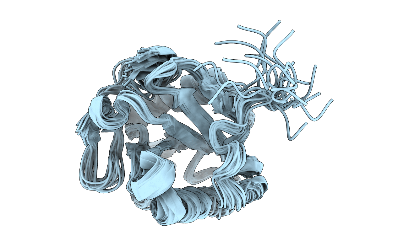
Conformers Submitted:
20
Macromolecular Entities
Polymer Type:polypeptide(L)
Molecule:TRYPAREDOXIN 1
Gene (Uniprot):TryX
Chain IDs:A
Chain Length:154
Number of Molecules:1
Biological Source:Crithidia fasciculata
Ligand Molecules
Primary Citation
NMR studies of the interaction of tryparedoxin with redox-inactive substrate homologues.
Biochemistry 42 14720 14728 (2003)
PMID: 14674746 DOI: 10.1021/bi030112d

Abstact

Tryparedoxins (TXNs) are trypanothione-dependent peroxiredoxin oxidoreductases involved in hydroperoxide detoxification that have been shown to determine virulence in trypanosomatids. The structure of (15)N,(13)C-doubly-labeled, C-terminally-His-tagged tryparedoxin 1 from Crithidia fasciculata (Cf TXN1) was elucidated by three-dimensional NMR spectroscopy. Global folding was found to be similar to the crystal structure, but regions near the active site, especially the onset of helix alpha1 with the redox-active Cys 43 and helix alpha2 relevant to substrate binding, were less well defined in solution. The redox-inactive inhibitory substrate analogue N(1),N(8)-bis(ophthalmyl)spermidine was used to study the substrate/TXN interaction by two-dimensional (1)H,(15)N NMR spectroscopy. The NMR data complemented by molecular modeling revealed several alternative modes of ligand binding. The results confirm and extend the concept of TXN action and specificity derived from X-ray analysis and site-directed mutagenesis and thus improve the rational basis for inhibitor design.

Legend

Protein

Chemical

Disease

Primary Citation of related structures