Deposition Date
2003-07-21
Release Date
2004-04-05
Last Version Date
2023-12-13
Entry Detail
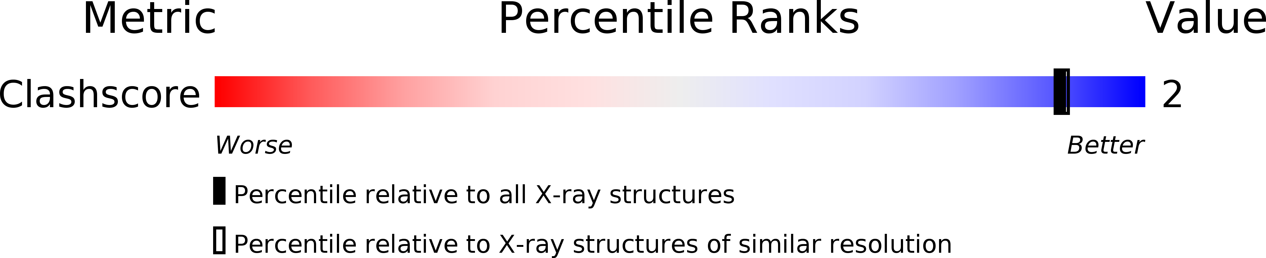
PDB ID:
1OKB
Keywords:
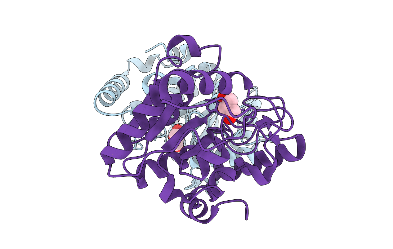
Title:
crystal structure of Uracil-DNA glycosylase from Atlantic cod (Gadus morhua)
Biological Source:
Source Organism(s):
GADUS MORHUA (Taxon ID: 8049)
Expression System(s):
Method Details:
Experimental Method:
Resolution:
1.90 Å
R-Value Free:
0.20
R-Value Work:
0.18
R-Value Observed:
0.18
Space Group:
P 1 21 1