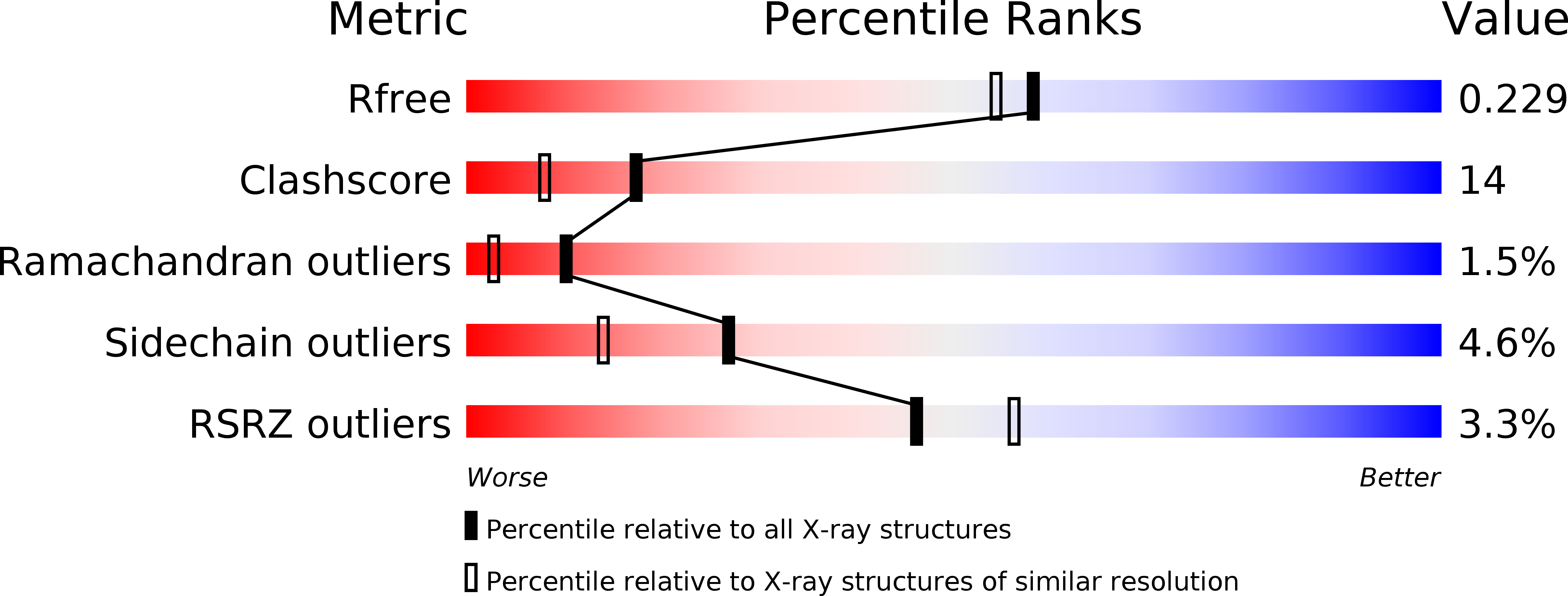
1OJ6 image
Deposition Date 2003-07-03
Release Date 2003-09-11
Last Version Date 2024-05-08
Entry Detail
PDB ID:
1OJ6
Title:
Human brain neuroglobin three-dimensional structure
Biological Source:
Source Organism:
Homo sapiens (Taxon ID: 9606)
Method Details:
Experimental Method:
Resolution:
1.95 Å
R-Value Free:
0.23
R-Value Work:
0.17
R-Value Observed:
0.17
Space Group:
P 1 21 1
Macromolecular Entities
Structures with similar UniProt ID
Protein Blast
Polymer Type:polypeptide(L)
Molecule:Neuroglobin
Gene (Uniprot):NGB
Mutagens:YES
Chain IDs:A, B, C, D
Chain Length:151
Number of Molecules:4
Biological Source:Homo sapiens
Primary Citation
Human Brain Neuroglobin Structure Reveals a Distinct Mode of Controlling Oxygen Affinity
Structure 11 1087 ? (2003)
PMID: 12962627 DOI: 10.1016/S0969-2126(03)00166-7

Abstact

Neuroglobin, mainly expressed in vertebrate brain and retina, is a recently identified member of the globin superfamily. Augmenting O(2) supply, neuroglobin promotes survival of neurons upon hypoxic injury, potentially limiting brain damage. In the absence of exogenous ligands, neuroglobin displays a hexacoordinated heme. O(2) and CO bind to the heme iron, displacing the endogenous HisE7 heme distal ligand. Hexacoordinated human neuroglobin displays a classical globin fold adapted to host the reversible bis-histidyl heme complex and an elongated protein matrix cavity, held to facilitate O(2) diffusion to the heme. The neuroglobin structure suggests that the classical globin fold is endowed with striking adaptability, indicating that hemoglobin and myoglobin are just two examples within a wide and functionally diversified protein homology superfamily.

Legend

Protein

Chemical

Disease

Primary Citation of related structures