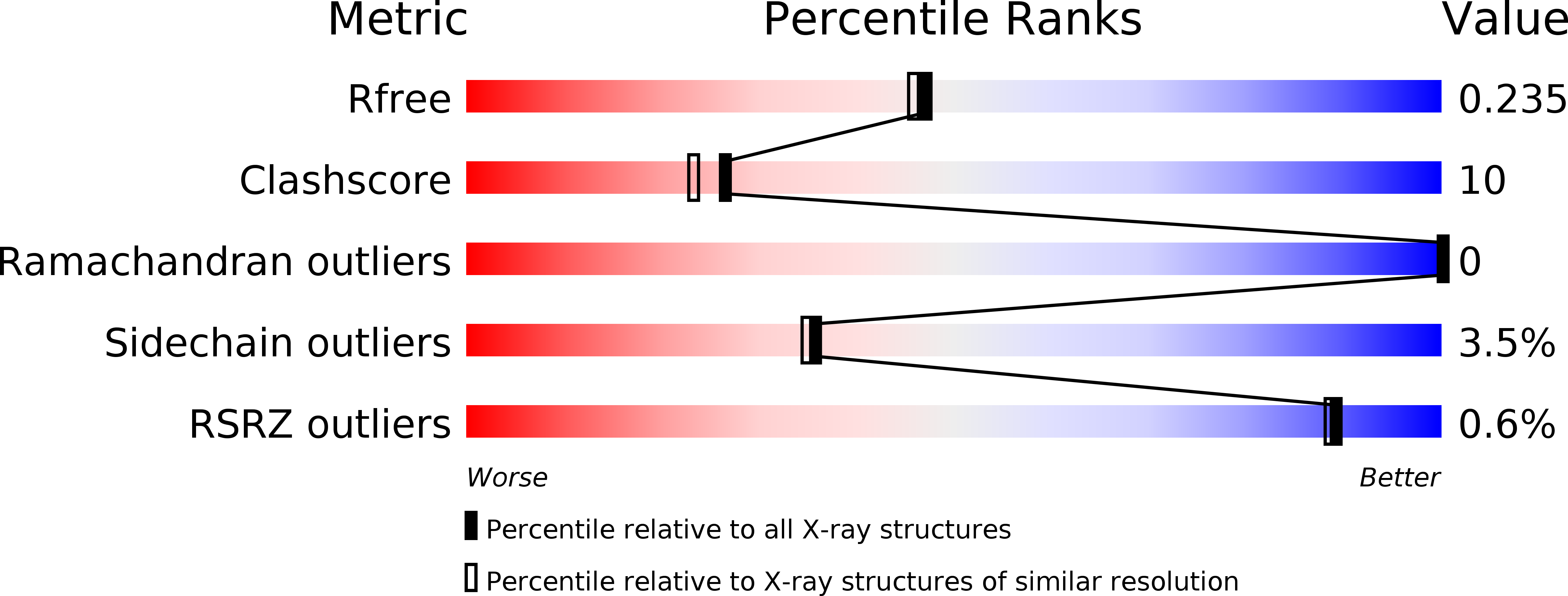
1OHT image
Deposition Date 2003-05-31
Release Date 2003-07-18
Last Version Date 2024-11-13
Entry Detail
PDB ID:
1OHT
Keywords:
Title:
Peptidoglycan recognition protein LB
Biological Source:
Source Organism:
Host Organism:
Method Details:
Experimental Method:
Resolution:
2.00 Å
R-Value Free:
0.24
R-Value Work:
0.21
R-Value Observed:
0.21
Space Group:
P 61 2 2
Macromolecular Entities
Polymer Type:polypeptide(L)
Molecule:CG14704 PROTEIN
Chain IDs:A
Chain Length:215
Number of Molecules:1
Biological Source:DROSOPHILA MELANOGASTER
Modified Residue
Compound ID Chain ID Parent Comp ID Details 2D Image
MSE A MET SELENOMETHIONINE
Primary Citation
Crystal Structure of Peptidoglycan Recognition Protein Lb from Drosophila Melanogaster
Nat.Immunol. 4 787 ? (2003)
PMID: 12845326 DOI: 10.1038/NI952

Abstact

The family of peptidoglycan recognition proteins (PGRPs) are associated with the recognition of the peptidoglycan of microbes and subsequent activation of signaling pathways for immune response. Here the crystal structure of Drosophila PGRP-LB is determined at a resolution of 2.0 A and shows an active-site cleft with a zinc cage. Poor conservation of surface residues at the cleft predicts a widely varying individual specificity of PGRPs for molecular patterns on microbial cell walls. At the back of this cleft is a putatively conserved distinctive groove. The location and mainly hydrophobic nature of the groove indicate that the back face serves for subsequent signaling after clustering of PGRP molecules by binding to polymeric cell wall components.

Legend

Protein

Chemical

Disease

Primary Citation of related structures