Deposition Date
2003-04-30
Release Date
2003-09-01
Last Version Date
2024-05-08
Entry Detail
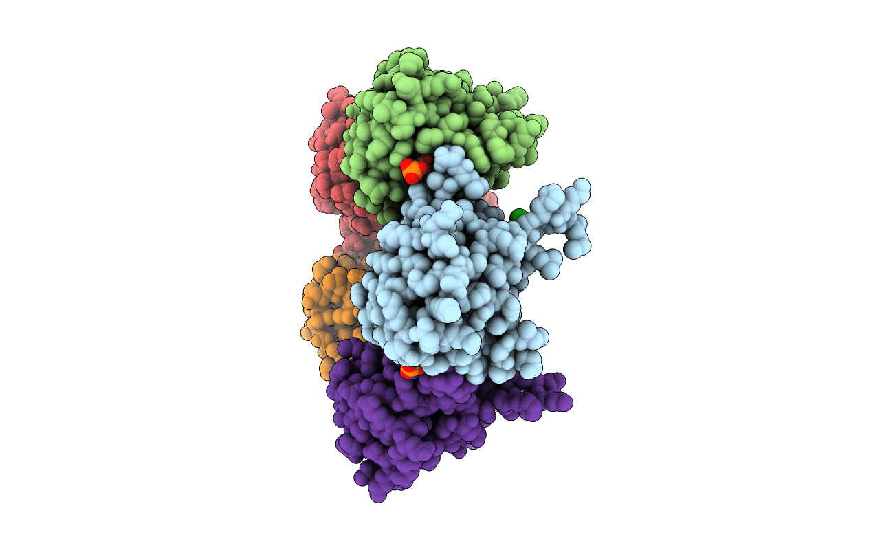
PDB ID:
1OGE
Keywords:
Title:
The Structure of Bacillus subtilis RbsD complexed with Ribose 5-phosphate
Biological Source:
Source Organism(s):
BACILLUS SUBTILIS (Taxon ID: 1423)
Method Details:
Experimental Method:
Resolution:
2.05 Å
R-Value Free:
0.22
R-Value Work:
0.19
R-Value Observed:
0.19
Space Group:
C 1 2 1