Deposition Date
2003-04-23
Release Date
2003-08-28
Last Version Date
2024-10-16
Entry Detail
PDB ID:
1OG1
Keywords:
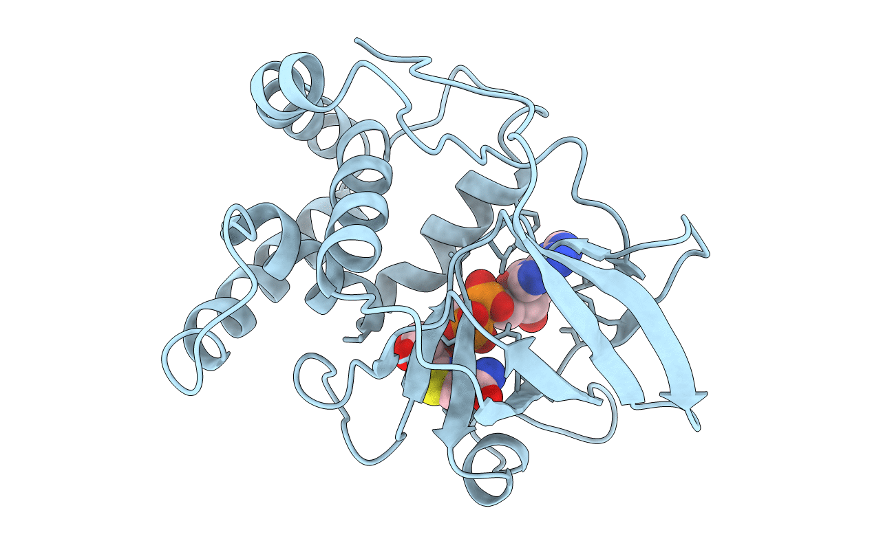
Title:
CRYSTAL STRUCTURE OF THE EUCARYOTIC MONO-ADP-RIBOSYLTRANSFERASE ART2.2 IN COMPLEX WITH TAD
Biological Source:
Source Organism(s):
RATTUS NORVEGICUS (Taxon ID: 10116)
Expression System(s):
Method Details:
Experimental Method:
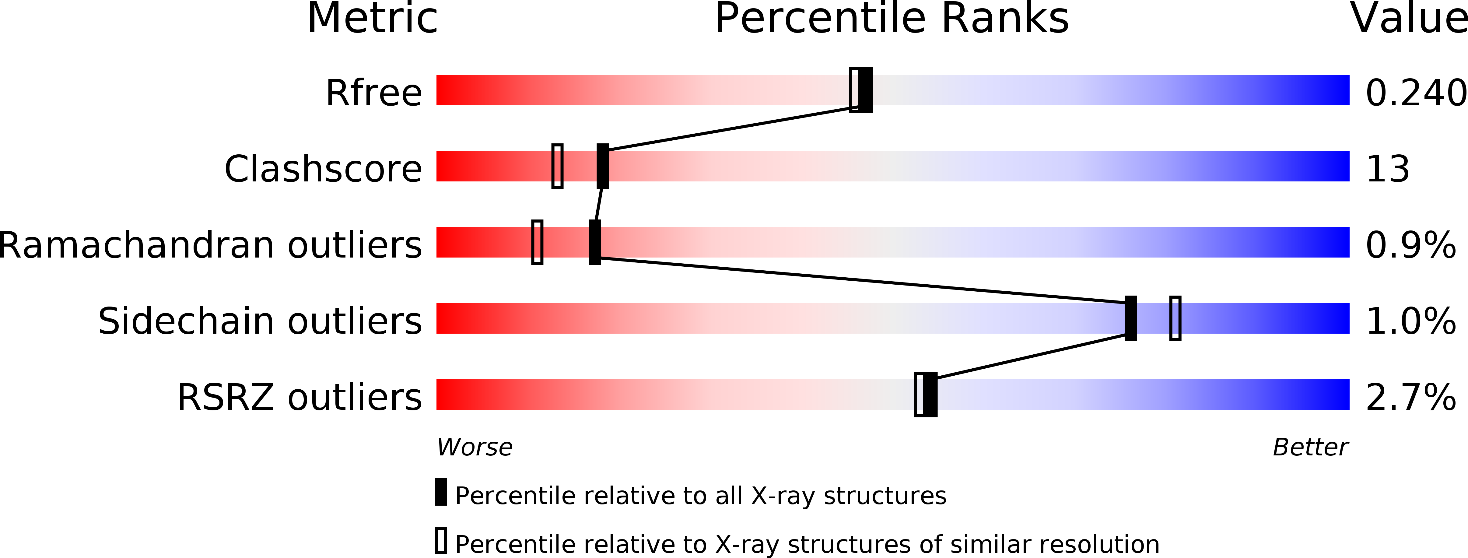
Resolution:
2.00 Å
R-Value Free:
0.24
R-Value Work:
0.20
R-Value Observed:
0.20
Space Group:
P 31 2 1