
Deposition Date
2003-04-22
Release Date
2003-09-18
Last Version Date
2024-11-13
Entry Detail
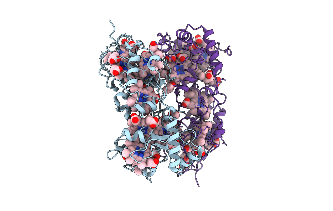
PDB ID:
1OFY
Keywords:
Title:
three dimensional structure of the reduced form of nine-heme cytochrome c at ph 7.5
Biological Source:
Source Organism(s):
DESULFOVIBRIO DESULFURICANS (Taxon ID: 876)
Method Details:
Experimental Method:
Resolution:
2.00 Å
R-Value Free:
0.27
R-Value Observed:
0.21
Space Group:
P 1 21 1
