Deposition Date
2003-04-21
Release Date
2003-06-19
Last Version Date
2024-05-08
Entry Detail
PDB ID:
1OFT
Keywords:
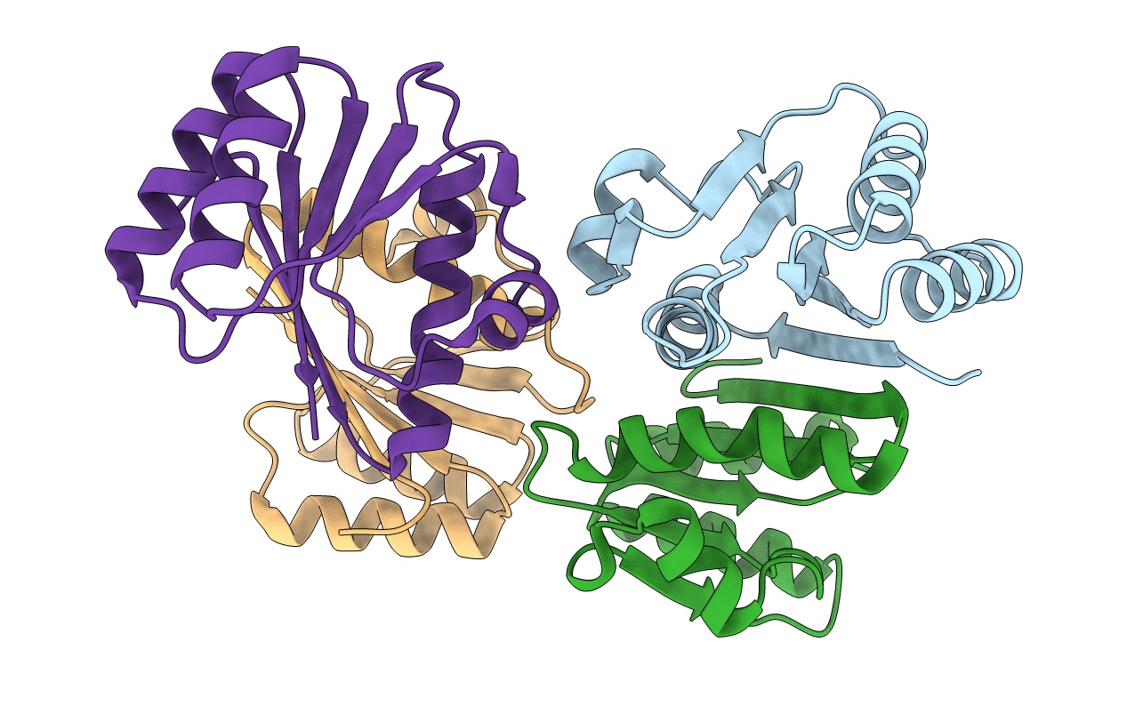
Title:
Crystal structure of SulA from Pseudomonas aeruginosa
Biological Source:
Source Organism(s):
PSEUDOMONAS AERUGINOSA (Taxon ID: 287)
Expression System(s):
Method Details:
Experimental Method:
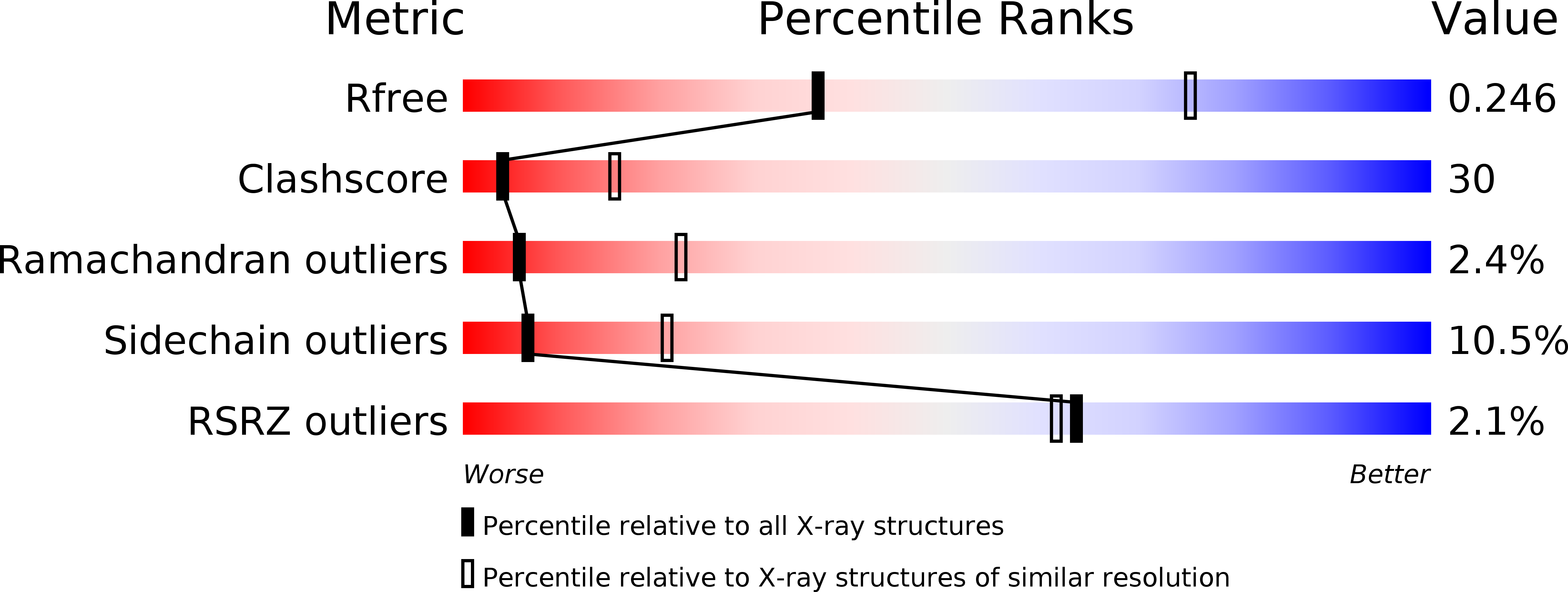
Resolution:
2.90 Å
R-Value Free:
0.28
R-Value Work:
0.24
R-Value Observed:
0.24
Space Group:
P 21 21 21