
Deposition Date
2003-04-15
Release Date
2004-04-19
Last Version Date
2024-11-13
Entry Detail
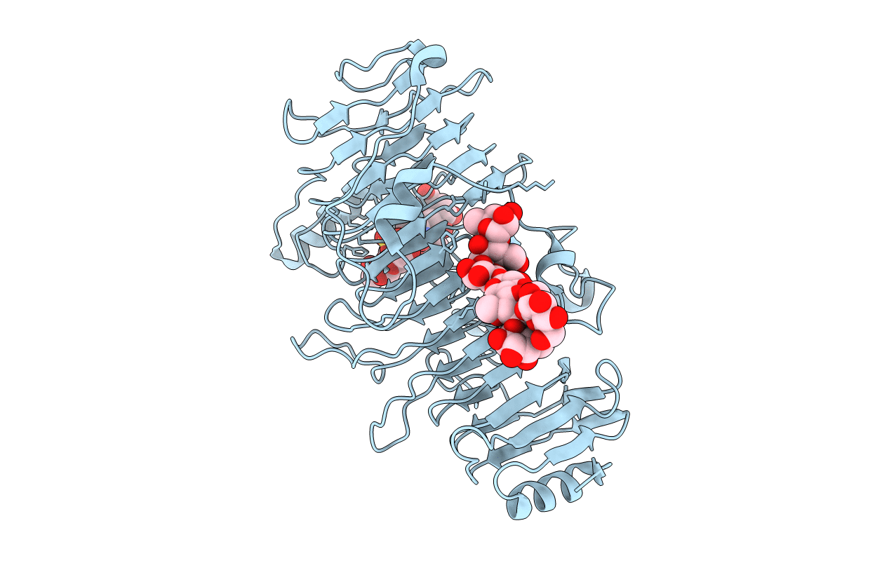
PDB ID:
1OFM
Keywords:
Title:
CRYSTAL STRUCTURE OF CHONDROITINASE B COMPLEXED TO CHONDROITIN 4-SULFATE TETRASACCHARIDE
Biological Source:
Source Organism(s):
PEDOBACTER HEPARINUS (Taxon ID: 984)
Expression System(s):
Method Details:
Experimental Method:
Resolution:
1.80 Å
R-Value Free:
0.18
R-Value Work:
0.14
R-Value Observed:
0.14
Space Group:
P 1 21 1
