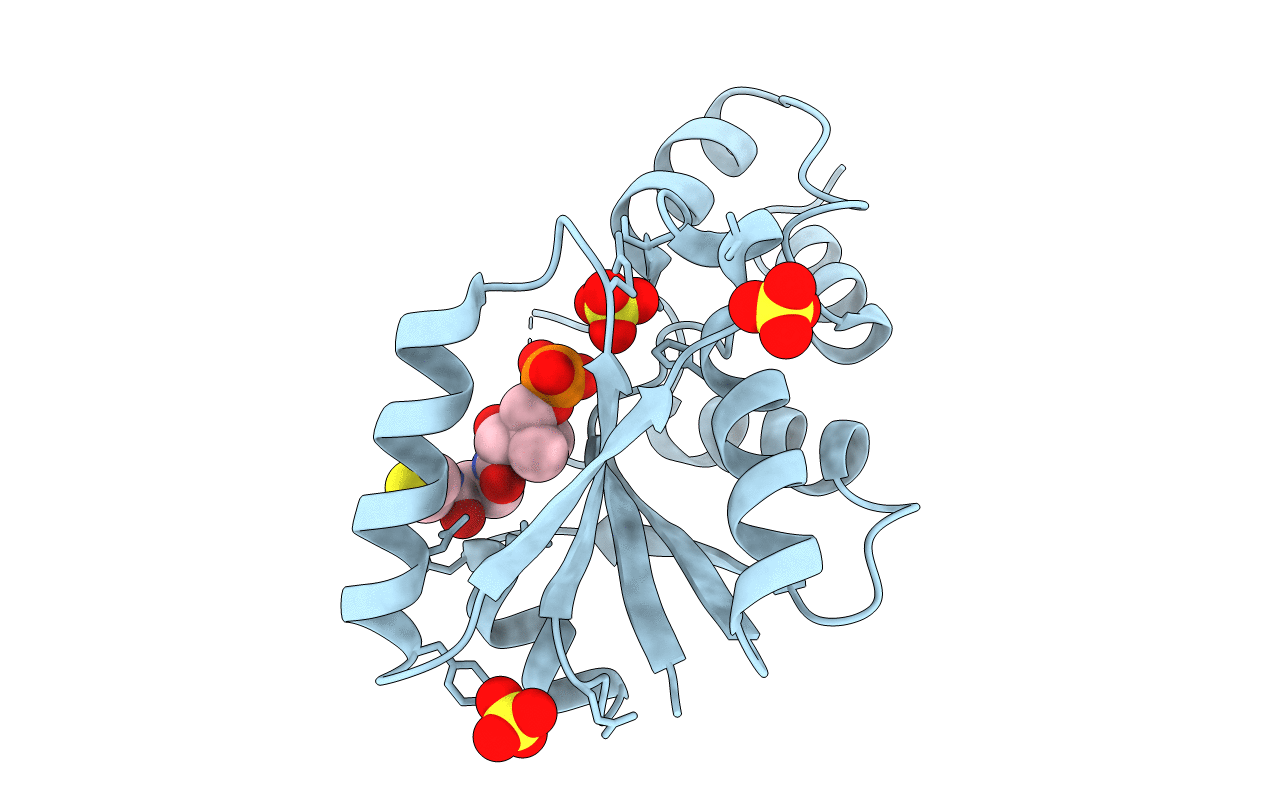
1OD6 image
Entry Detail
PDB ID:
1OD6
Keywords:
Title:
The Crystal Structure of Phosphopantetheine adenylyltransferase from Thermus Thermophilus in complex with 4'-phosphopantetheine
Biological Source:
Source Organism:
Host Organism:
PDB Version:
Deposition Date:
2003-02-13
Release Date:
2003-03-13
Method Details:
Experimental Method:
Resolution:
1.50 Å
R-Value Free:
0.20
R-Value Work:
0.19
R-Value Observed:
0.19
Space Group:
H 3 2
Macromolecular Entities
Polymer Type:polypeptide(L)
Description:PHOSPHOPANTETHEINE ADENYLYLTRANSFERASE
Chain IDs:A
Chain Length:160
Number of Molecules:1
Biological Source:THERMUS THERMOPHILUS
Primary Citation
Structure and Implications for the Thermal Stability of Phosphopantetheine Adenylyltransferase from Thermus Thermophilus.
Acta Crystallogr.,Sect.D 60 97 ? (2004)
PMID: 14684898 DOI: 10.1107/S0907444903025319

Abstact

Phosphopantetheine adenylyltransferase (PPAT) is an essential enzyme in bacteria that catalyzes the rate-limiting step in coenzyme A (CoA) biosynthesis by transferring an adenylyl group from ATP to 4'-phosphopantetheine (Ppant), yielding 3'-dephospho-CoA (dPCoA). The crystal structure of PPAT from Thermus thermophilus HB8 (Tt PPAT) complexed with Ppant has been determined by the molecular-replacement method at 1.5 A resolution. The overall fold of the enzyme is almost the same as that of Escherichia coli PPAT, a hexamer having point group 32. The asymmetric unit of Tt PPAT contains a monomer and the crystallographic triad and dyad coincide with the threefold and twofold axes of the hexamer, respectively. Most of the important atoms surrounding the active site in E. coli PPAT are conserved in Tt PPAT, indicating similarities in their substrate binding and enzymatic reaction. The notable difference between E. coli PPAT and Tt PPAT is the simultaneous substrate recognition by all six subunits of Tt PPAT compared with substrate recognition by only three subunits in E. coli PPAT. Comparative analysis also revealed that the higher stability of Tt PPAT arises from stabilization of each subunit by hydrophobic effects, hydrogen bonds and entropic effects.

Legend

Protein

Chemical

Disease

Primary Citation of related structures