1OCY image
Deposition Date 2003-02-11
Release Date 2003-07-24
Last Version Date 2024-05-08
Entry Detail
PDB ID:
1OCY
Title:
Structure of the receptor-binding domain of the bacteriophage T4 short tail fibre
Biological Source:
Source Organism:
Host Organism:
Method Details:
Experimental Method:
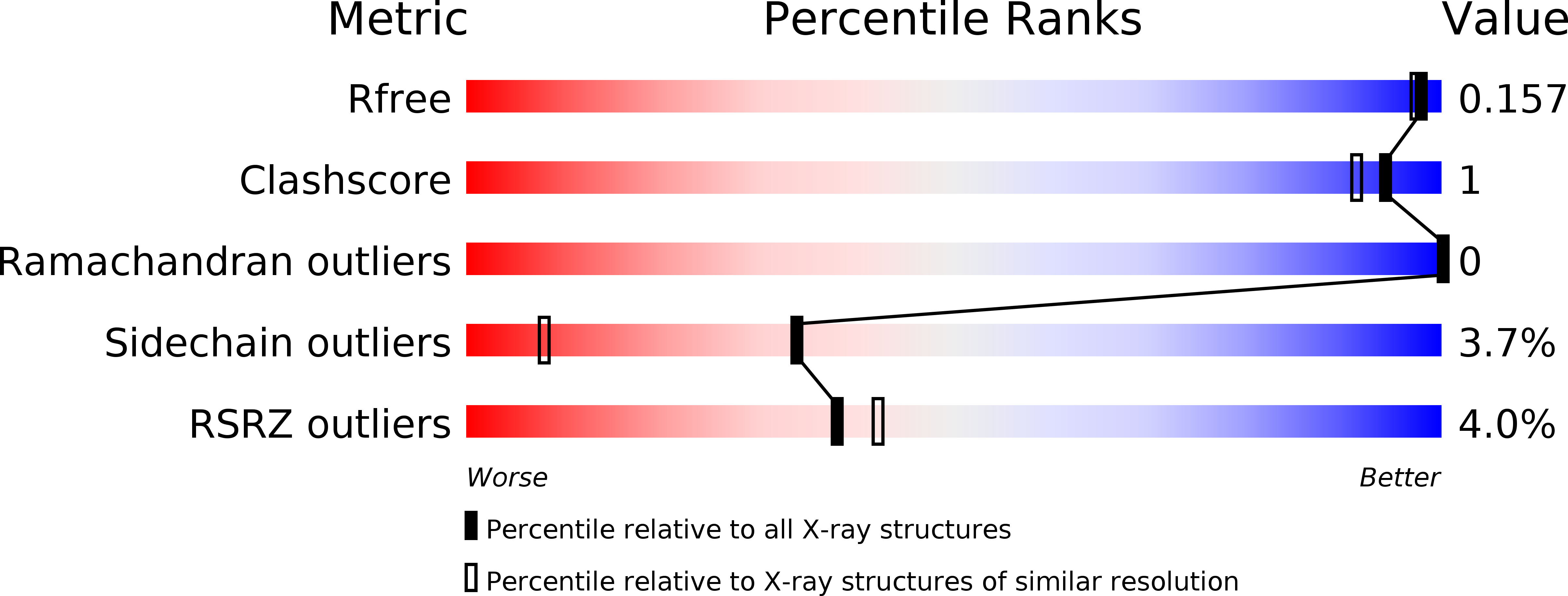
Resolution:
1.50 Å
R-Value Free:
0.15
R-Value Work:
0.14
Space Group:
P 3 2 1
Macromolecular Entities
Structures with similar UniProt ID
Protein Blast
Polymer Type:polypeptide(L)
Molecule:BACTERIOPHAGE T4 SHORT TAIL FIBRE
Chain IDs:A
Chain Length:198
Number of Molecules:1
Biological Source:BACTERIOPHAGE T4
Primary Citation
The Structure of the Receptor-Binding Domain of the Bacteriophage T4 Short Tail Fibre Reveals a Knitted Trimeric Metal-Binding Fold
J.Mol.Biol. 331 361 373 (2003)
PMID: 12888344 DOI: 10.1016/S0022-2836(03)00755-1

Abstact

Adsorption of T4 bacteriophage to the Escherichia coli host cell is mediated by six long and six short tail fibres. After at least three long tail fibres have bound, short tail fibres extend and bind irreversibly to the core region of the host cell lipo-polysaccharide (LPS), serving as inextensible stays during penetration of the cell envelope by the tail tube. The short tail fibres consist of a parallel, in-register, trimer of gene product 12 (gp12).X-ray crystallography at 1.5A resolution of a protease-stable fragment of gp12 generated in the presence of zinc chloride reveals the structure of the C-terminal receptor-binding domain. It has a novel "knitted" fold, consisting of three extensively intertwined monomers. It reveals a metal-binding site, containing a zinc ion coordinated by six histidine residues in an octahedral conformation. We also suggest an LPS-binding region.

Legend

Protein

Chemical

Disease

Primary Citation of related structures
Feedback Form
Name
Email
Institute
Feedback