1OCS image
Entry Detail
PDB ID:
1OCS
Keywords:
Title:
Crystal structure of the yeast PX-doamin protein Grd19p (sorting nexin3) complexed to phosphatidylinosytol-3-phosphate.
Biological Source:
Source Organism:
Host Organism:
PDB Version:
Deposition Date:
2003-02-10
Release Date:
2003-12-12
Method Details:
Experimental Method:
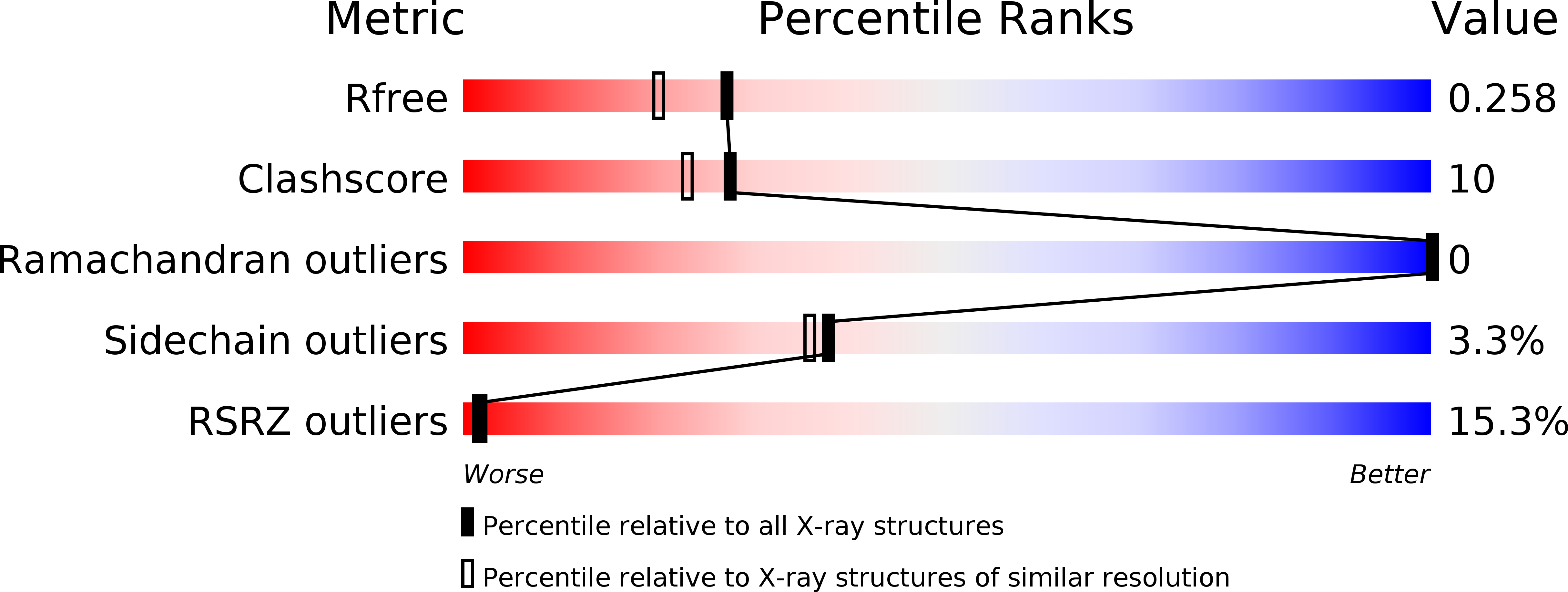
Resolution:
2.03 Å
R-Value Free:
0.24
R-Value Work:
0.21
R-Value Observed:
0.21
Space Group:
P 61 2 2
Macromolecular Entities
Polymer Type:polypeptide(L)
Description:SORTING NEXIN GRD19
Chain IDs:A
Chain Length:162
Number of Molecules:1
Biological Source:SACCHAROMYCES CEREVISIAE
Modified Residue
Compound ID Chain ID Parent Comp ID Details 2D Image
CME A CYS S,S-(2-HYDROXYETHYL)THIOCYSTEINE
Ligand Molecules
Primary Citation
Crystal Structure of the Yeast Phox Homology (Px) Protein Grd19P (Sorting Nexin 3) Complexed to Phosphatidylinositol-3-Phosphate
J.Biol.Chem. 278 50371 ? (2003)
PMID: 14514667 DOI: 10.1074/JBC.M304392200

Abstact

Phox homology (PX) domains have been recently identified in a number of different proteins and are involved in various cellular functions such as vacuolar targeting and membrane protein trafficking. It was shown that these modules of about 130 amino acids specifically binding to phosphoinositides and that this interaction is crucial for their cellular function. The yeast genome contains 17 PX domain proteins. One of these, Grd19p, is involved in the localization of the late Golgi membrane proteins DPAP A and Kex2p. Grd19p consists of the PX domain with 30 extra residues at the N-terminal and is homologous to the functionally characterized human sorting nexin protein SNX3. We determined the 2.0 A crystal structure of Grd19p in the free form and in complex with d-myo-phosphatidylinositol 3-phosphate (diC4PtdIns(3)P), representing the first case of both free and ligand-bound conformations of the same PX module. The ligand occupies a well defined positively charged binding pocket at the interface between the beta-sheet and alpha-helical parts of the molecule. The structure of the free and bound protein are globally similar but show some significant differences in a region containing a polyproline peptide and a putative membrane attachment site.

Legend

Protein

Chemical

Disease

Primary Citation of related structures