1OBW image
Deposition Date 1996-10-09
Release Date 1997-09-04
Last Version Date 2024-04-03
Entry Detail
PDB ID:
1OBW
Keywords:
Title:
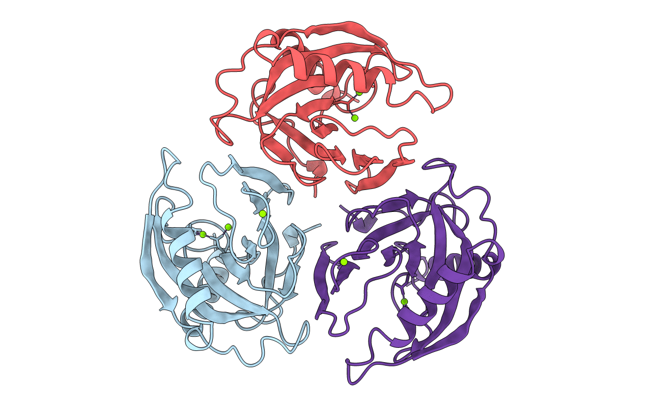
STRUCTURE OF INORGANIC PYROPHOSPHATASE
Biological Source:
Source Organism:
Escherichia coli (Taxon ID: 562)
Host Organism:
Method Details:
Experimental Method:
Resolution:
1.90 Å
R-Value Free:
0.22
R-Value Work:
0.17
Space Group:
P 32 2 1
Macromolecular Entities
Polymer Type:polypeptide(L)
Molecule:INORGANIC PYROPHOSPHATASE
Gene (Uniprot):ppa
Chain IDs:A, B, C
Chain Length:175
Number of Molecules:3
Biological Source:Escherichia coli
Ligand Molecules
Primary Citation
Crystal structure of holo inorganic pyrophosphatase from Escherichia coli at 1.9 A resolution. Mechanism of hydrolysis.
Biochemistry 36 7754 7760 (1997)
PMID: 9201917 DOI: 10.1021/bi962637u

Abstact

Crystalline holo inorganic pyrophosphatase from Escherichia coli was grown in the presence of 250 mM MgCl2. The crystal structure has been solved by Patterson search techniques and refined to an R-factor of 17.6% at 1.9 A resolution. The upper estimate of the root-mean-square error in atomic positions is 0.26 A. These crystals belong to space group P3(2)21 with unit cell dimensions a = b = 110.27 A and c = 78.17 A. The asymmetric unit contains a trimer of subunits, i.e., half of the hexameric molecule. In the central cavity of the enzyme molecule, three Mg2+ ions, each shared by two subunits of the hexamer, are found. In the active sites of two crystallographically independent subunits, two Mg2+ ions are bound. The second active site Mg2+ ion is missing in the third subunit. A mechanism of catalysis is proposed whereby a water molecule activated by a Mg2+ ion and Tyr 55 play essential roles.

Legend

Protein

Chemical

Disease

Primary Citation of related structures