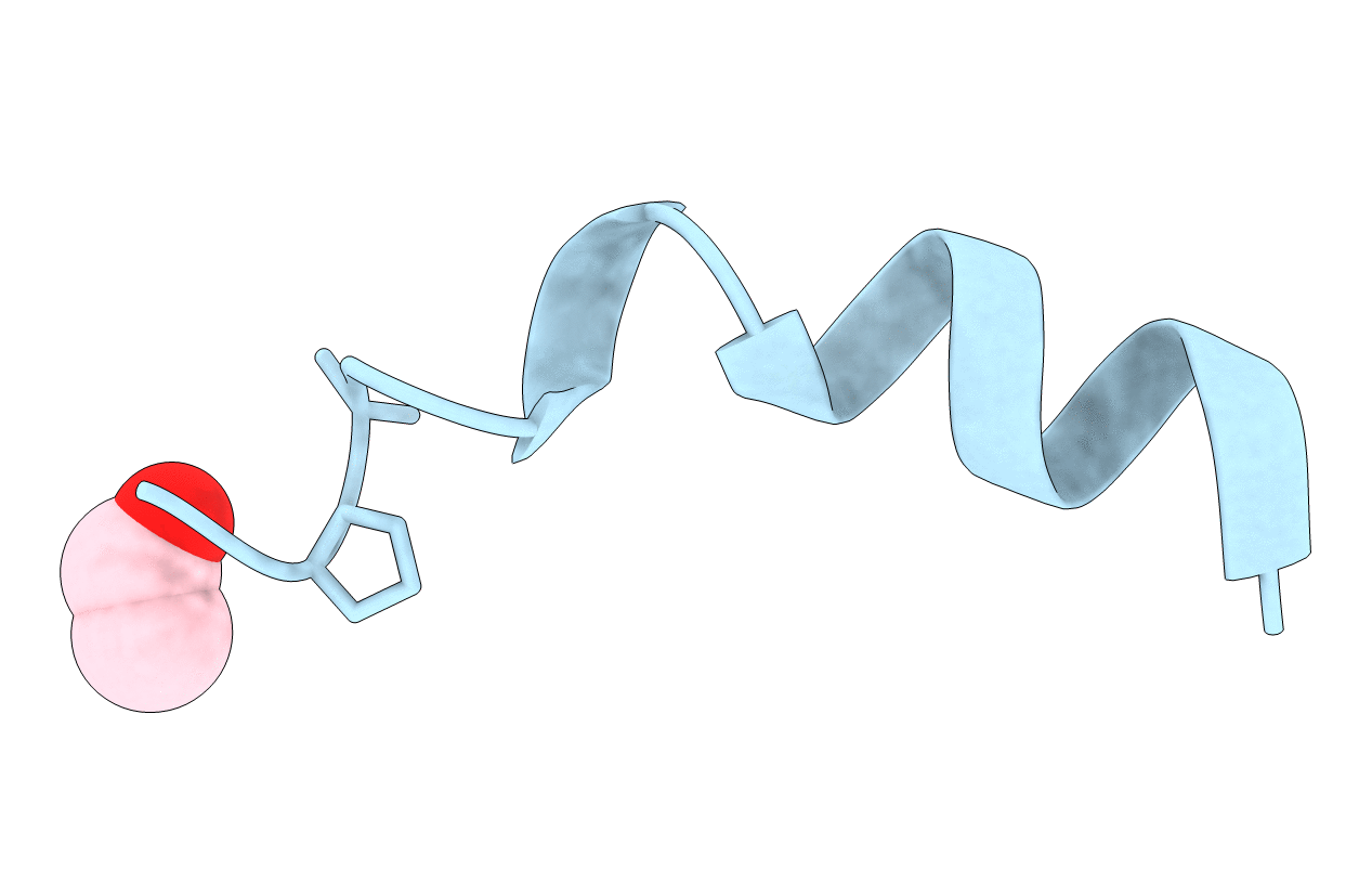
1OB4 image
Entry Detail
PDB ID:
1OB4
Keywords:
Title:
Cephaibol A
Biological Source:
Source Organism:
PDB Version:
Deposition Date:
2003-01-24
Release Date:
2003-12-11
Method Details:
Experimental Method:
Resolution:
0.95 Å
R-Value Free:
0.11
R-Value Observed:
0.08
Space Group:
P 21 21 2
Macromolecular Entities
Polymer Type:polypeptide(L)
Description:CEPHAIBOL A
Chain IDs:A
Chain Length:17
Number of Molecules:1
Biological Source:ACREMONIUM TUBAKII
Modified Residue
Compound ID Chain ID Parent Comp ID Details 2D Image
AIB A ALA ALPHA-AMINOISOBUTYRIC ACID
HYP A PRO 4-HYDROXYPROLINE
PHL A PHE L-PHENYLALANINOL
Ligand Molecules
Peptide-like Molecules
PRD_000156
Primary Citation
Crystal Structures of Cephaibols
J.Pept.Sci. 9 745 ? (2003)
PMID: 14658793 DOI: 10.1002/PSC.496

Abstact

The crystal structures of the peptaibol antibiotics cephaibol A, cephaibol B and cephaibol C have been determined at ca. 0.9 A resolution. All three adopt a helical conformation with a sharp bend (of about 55 degrees) at the central hydroxyproline. All isovalines were found to possess the D configuration, superposition of all four models (there are two independent molecules in the cephaibol B structure) shows that the N-terminal helix is rigid and the C-terminus is flexible. There are differences in the hydrogen bonding patterns for the three structures that crystallize in different space groups despite relatively similar unit cell dimensions, but only in the case of cephaibol C does the packing emulate the formation of a membrane channel believed to be important for their biological function.

Legend

Protein

Chemical

Disease

Primary Citation of related structures