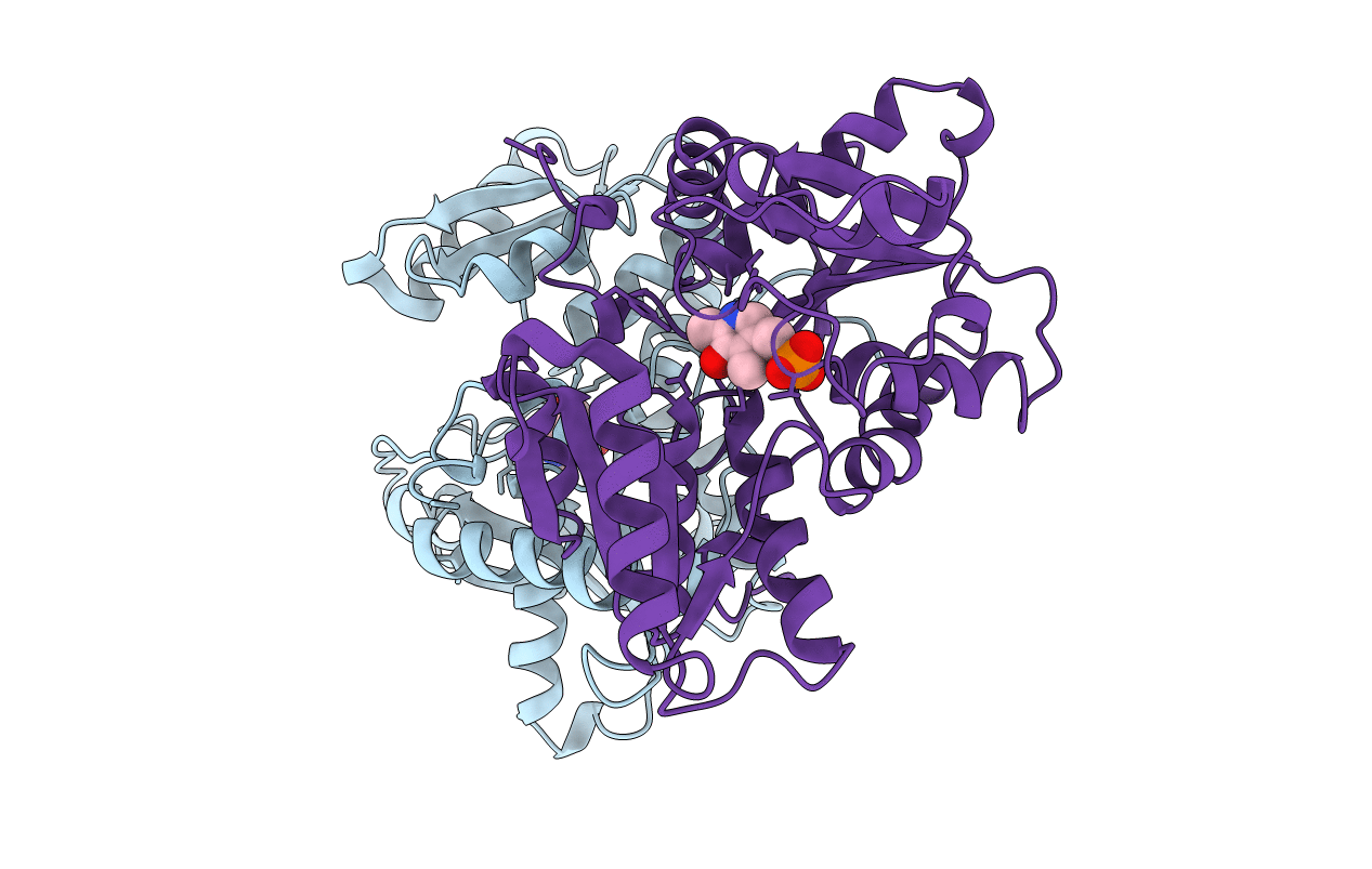
1OAS image
Entry Detail
PDB ID:
1OAS
Keywords:
Title:
O-ACETYLSERINE SULFHYDRYLASE FROM SALMONELLA TYPHIMURIUM
Biological Source:
Source Organism:
Host Organism:
PDB Version:
Deposition Date:
1999-01-29
Release Date:
2000-01-28
Method Details:
Experimental Method:
Resolution:
2.20 Å
R-Value Free:
0.20
R-Value Work:
0.17
Space Group:
P 21 21 21
Macromolecular Entities
Polymer Type:polypeptide(L)
Description:O-ACETYLSERINE SULFHYDRYLASE
Chain IDs:A, B
Chain Length:322
Number of Molecules:2
Biological Source:Salmonella typhimurium
Ligand Molecules
Primary Citation
Three-dimensional structure of O-acetylserine sulfhydrylase from Salmonella typhimurium.
J.Mol.Biol. 283 121 133 (1998)
PMID: 9761678 DOI: 10.1006/jmbi.1998.2037

Abstact

The last step in cysteine biosynthesis in enteric bacteria is catalyzed by the pyridoxal 5'-phosphate-dependent enzyme O-acetylserine sulfhydrylase. Here we report the crystal structure at 2.2 A resolution of the A-isozyme of O-acetylserine sulfhydrylase isolated from Salmonella typhimurium. O-acetylserine sulfhydrylase shares the same fold with tryptophan synthase-beta from Salmonella typhimurium but the sequence identity level is below 20%. There are some major structural differences: the loops providing the interface to the alpha-subunit in tryptophan synthase-beta and two surface helices of tryptophan synthase-beta are missing in O-acetylserine sulfhydrylase. The hydrophobic channel for indole transport from the alpha to the beta active site of tryptophan synthase-beta is, not unexpectedly, also absent in O-acetylserine sulfhydrylase. The dimer interface, on the other hand, is more or less conserved in the two enzymes. The active site cleft of O-acetylserine sulfhydrylase is wider and therefore more exposed to the solvent. A possible binding site for the substrate O-acetylserine is discussed.

Legend

Protein

Chemical

Disease

Primary Citation of related structures