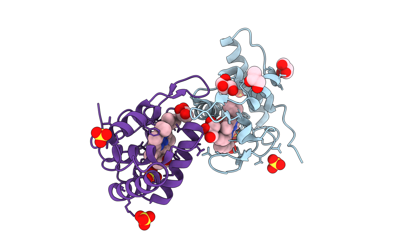
1OAE image
Entry Detail
PDB ID:
1OAE
Title:
Crystal structure of the reduced form of cytochrome c" from Methylophilus methylotrophus
Biological Source:
PDB Version:
Deposition Date:
2003-01-09
Release Date:
2004-03-26
Method Details:
Experimental Method:
Resolution:
1.95 Å
R-Value Free:
0.24
R-Value Work:
0.18
R-Value Observed:
0.18
Space Group:
P 21 21 21
Macromolecular Entities
Polymer Type:polypeptide(L)
Description:CYTOCHROME C"
Chain IDs:A, B
Chain Length:124
Number of Molecules:2
Biological Source:METHYLOPHILUS METHYLOTROPHUS
Primary Citation
Structural Evidence for a Proton Transfer Pathway Coupled with Haem Reduction of Cytochrome C" from Methylophilus Methylotrophus.
J.Biol.Inorg.Chem. 11 189 ? (2006)
PMID: 16341897 DOI: 10.1007/S00775-005-0065-6

Abstact

The crystal structures of the oxidized and reduced forms of cytochrome c" from Methylophilus methylotrophus were solved from X-ray synchrotron data to atomic resolution. The overall fold of the molecule in the two redox states is very similar and is comparable to that of the oxygen-binding protein from the purple phototrophic bacterium Rhodobacter sphaeroides. However, significant modifications occur near the haem group, in particular the detachment from axial binding of His95 observed upon reduction as well as the adoption of different conformations of some protonatable residues that form a possible proton path from the haem pocket to the protein surface. These changes are associated with the previously well characterized redox-Bohr behaviour of this protein. Furthermore they provide a model for one of the presently proposed mechanisms of proton translocation in the much more complex protein cytochrome c oxidase.

Legend

Protein

Chemical

Disease

Primary Citation of related structures