1OAC image
Deposition Date 1995-09-27
Release Date 1996-04-03
Last Version Date 2025-03-26
Entry Detail
PDB ID:
1OAC
Keywords:
Title:
CRYSTAL STRUCTURE OF A QUINOENZYME: COPPER AMINE OXIDASE OF ESCHERICHIA COLI AT 2 ANGSTROEMS RESOLUTION
Biological Source:
Source Organism:
Escherichia coli (Taxon ID: 562)
Method Details:
Experimental Method:
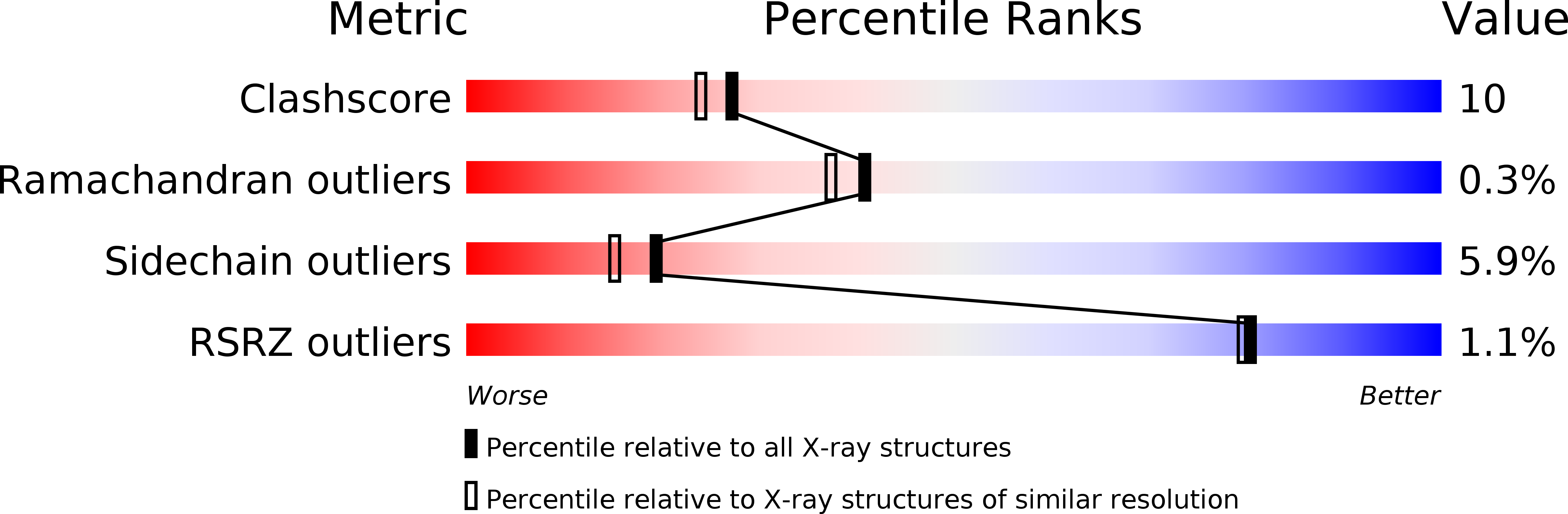
Resolution:
2.00 Å
R-Value Observed:
0.16
Space Group:
P 21 21 21
Macromolecular Entities
Polymer Type:polypeptide(L)
Molecule:COPPER AMINE OXIDASE
Gene (Uniprot):tynA
Chain IDs:A, B
Chain Length:727
Number of Molecules:2
Biological Source:Escherichia coli
Modified Residue
Compound ID Chain ID Parent Comp ID Details 2D Image
TPQ A TYR ?
Primary Citation
Crystal structure of a quinoenzyme: copper amine oxidase of Escherichia coli at 2 A resolution.
Structure 3 1171 1184 (1995)
PMID: 8591028 DOI: 10.1016/S0969-2126(01)00253-2

Abstact

BACKGROUND Copper amine oxidases are a ubiquitous and novel group of quinoenzymes that catalyze the oxidative deamination of primary amines to the corresponding aldehydes, with concomitant reduction of molecular oxygen to hydrogen peroxide. The enzymes are dimers of identical 70-90 kDa subunits, each of which contains a single copper ion and a covalently bound cofactor formed by the post-translational modification of a tyrosine side chain to 2,4,5-trihydroxyphenylalanine quinone (TPQ). RESULTS The crystal structure of amine oxidase from Escherichia coli has been determined in both an active and an inactive form. The only structural differences are in the active site, where differences in copper coordination geometry and in the position and interactions of the redox cofactor, TPQ, are observed. Each subunit of the mushroom-shaped dimer comprises four domains: a 440 amino acid C-terminal beta sandwich domain, which contains the active site and provides the dimer interface, and three smaller peripheral alpha/beta domains (D1-D3), each of about 100 amino acids. D2 and D3 show remarkable structural and sequence similarity to each other and are conserved throughout the quinoenzyme family. In contrast, D1 is absent from some amine oxidases. The active sites are well buried from solvent and lie some 35 A apart, connected by a pair of beta hairpin arms. CONCLUSIONS The crystal structure of E. coli copper amine oxidase reveals a number of unexpected features and provides a basis for investigating the intriguing similarities and differences in catalytic mechanism of members of this enzyme family. In addition to the three conserved histidines that bind the copper, our studies identify a number of other conserved residues close to the active site, including a candidate for the catalytic base and a fourth conserved histidine which is involved in an interesting intersubunit interaction.

Legend

Protein

Chemical

Disease

Primary Citation of related structures