1O9K image
Deposition Date 2002-12-16
Release Date 2003-03-06
Last Version Date 2024-10-23
Entry Detail
PDB ID:
1O9K
Keywords:
Title:
Crystal structure of the retinoblastoma tumour suppressor protein bound to E2F peptide
Biological Source:
Source Organism:
HOMO SAPIENS (Taxon ID: 9606)
Host Organism:
Method Details:
Experimental Method:
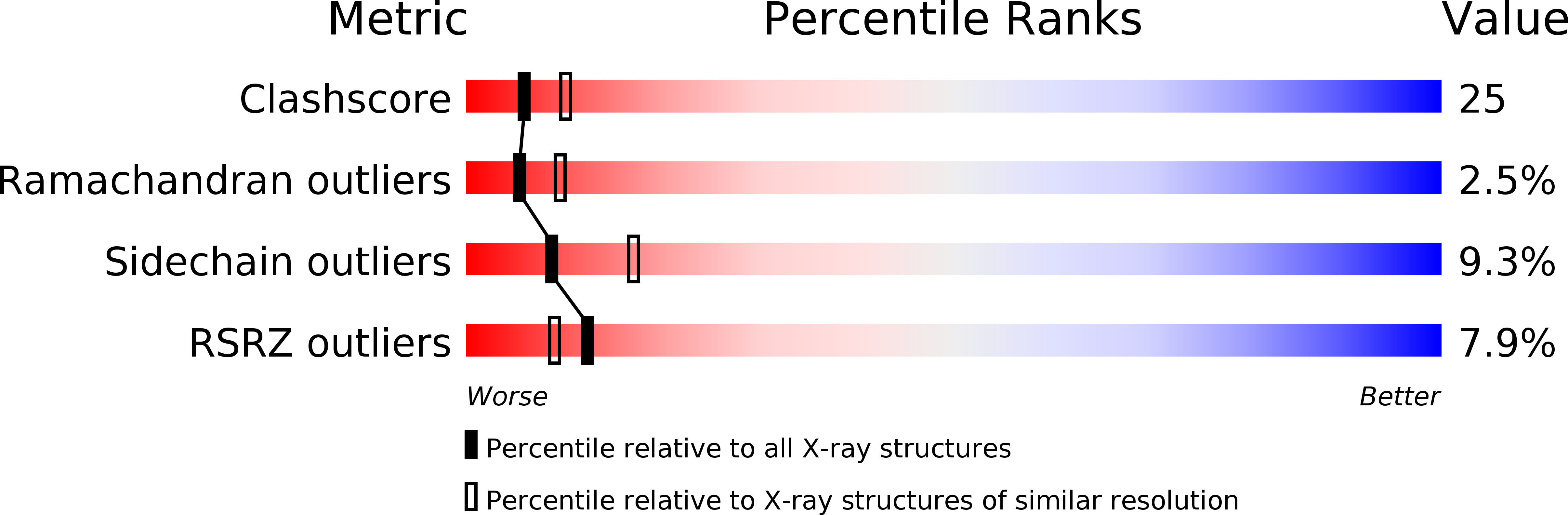
Resolution:
2.60 Å
R-Value Free:
0.28
R-Value Work:
0.22
R-Value Observed:
0.23
Space Group:
C 1 2 1
Macromolecular Entities
Structures with similar UniProt ID
Protein Blast
Polymer Type:polypeptide(L)
Molecule:RETINOBLASTOMA-ASSOCIATED PROTEIN
Gene (Uniprot):RB1
Chain IDs:A, C, E, G
Chain Length:218
Number of Molecules:4
Biological Source:HOMO SAPIENS
Structures with similar UniProt ID
Protein Blast
Polymer Type:polypeptide(L)
Molecule:RETINOBLASTOMA-ASSOCIATED PROTEIN
Gene (Uniprot):RB1
Chain IDs:B, D, F, H
Chain Length:152
Number of Molecules:4
Biological Source:HOMO SAPIENS
Structures with similar UniProt ID
Protein Blast
Polymer Type:polypeptide(L)
Molecule:TRANSCRIPTION FACTOR E2F1
Gene (Uniprot):E2F1
Chain IDs:I (auth: P), J (auth: Q), K (auth: R), L (auth: S)
Chain Length:18
Number of Molecules:4
Biological Source:HOMO SAPIENS
Primary Citation
Crystal Structure of the Retinoblastoma Tumor Suppressor Protein Bound to E2F and the Molecular Basis of its Regulation
Proc.Natl.Acad.Sci.USA 100 2363 ? (2003)
PMID: 12598654 DOI: 10.1073/PNAS.0436813100

Abstact

The retinoblastoma tumor suppressor protein (pRb) regulates the cell cycle, facilitates differentiation, and restrains apoptosis. Furthermore, dysfunctional pRb is thought to be involved in the development of most human malignancies. Many of the functions of pRb are mediated by its regulation of the E2F transcription factors. To understand the structural basis for this regulation, we have determined the crystal structure of a fragment of E2F in complex with the pocket domain of the tumor suppressor protein. The pRb pocket, comprising the A and B cyclin-like domains, is the major focus of tumourigenic mutations in the protein. The fragment of E2F used in our structural studies, residues 409-426 of E2F-1, represents the core of the pRb-binding region of the transcription factor. The structure shows that E2F binds at the interface of the A and B domains of the pocket making extensive interactions with conserved residues from both. We show by solution studies that a second site, probably contained within the "marked box" region of E2F, is responsible for additional interactions with the pRb pocket but is insufficient for complex formation on its own. In addition, we show that the interaction of the core binding fragment of E2F with pRb is inhibited by phosphorylation of the tumor suppressor protein by CDK2cyclin DE. Finally, our data reveal that the tight binding of the human papillomavirus E7 oncoprotein to pRb prevents subsequent interactions with the marked box region of E2F but not with its core binding region.

Legend

Protein

Chemical

Disease

Primary Citation of related structures