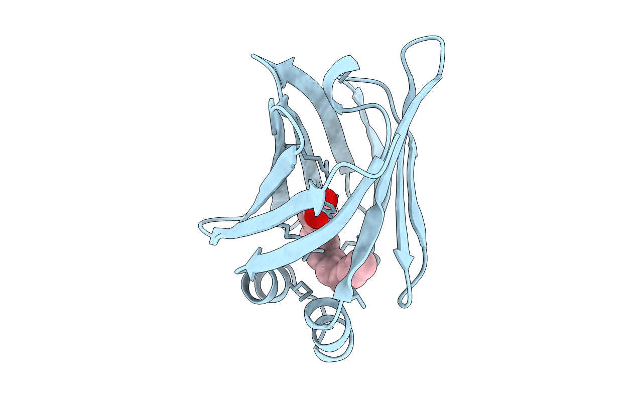
1O8V image
Deposition Date 2002-12-04
Release Date 2003-06-19
Last Version Date 2024-11-13
Entry Detail
PDB ID:
1O8V
Title:
The crystal structure of Echinococcus granulosus fatty-acid-binding protein 1
Biological Source:
Source Organism:
Host Organism:
Method Details:
Experimental Method:
Resolution:
1.60 Å
R-Value Free:
0.21
R-Value Work:
0.17
R-Value Observed:
0.17
Space Group:
P 1 21 1
Macromolecular Entities
Structures with similar UniProt ID
Protein Blast
Polymer Type:polypeptide(L)
Molecule:FATTY ACID BINDING PROTEIN HOMOLOG
Gene (Uniprot):FABP1
Chain IDs:A
Chain Length:134
Number of Molecules:1
Biological Source:ECHINOCOCCUS GRANULOSUS
Modified Residue
Compound ID Chain ID Parent Comp ID Details 2D Image
CSO A CYS S-HYDROXYCYSTEINE
Ligand Molecules
Primary Citation
The Crystal Structure of Echinococcus Granulosus Fatty-Acid-Binding Protein 1
Biochim.Biophys.Acta 1649 40 ? (2003)
PMID: 12818189 DOI: 10.1016/S1570-9639(03)00151-1

Abstact

We describe the 1.6 A crystal structure of the fatty-acid-binding protein EgFABP1 from the parasitic platyhelminth Echinococcus granulosus. E. granulosus causes hydatid disease, which is a major zoonosis. EgFABP1 has been implicated in the acquisition, storage, and transport of lipids, and may be important to the organism since it is incapable of synthesising most of its lipids de novo. Moreover, EgFABP1 is a promising candidate for a vaccine against hydatid disease. The crystal structure reveals that EgFABP1 has the expected 10-stranded beta-barrel fold typical of the family of intracellular lipid-binding proteins, and that it is structurally most similar to P2 myelin protein. We describe the comparison of the crystal structure of EgFABP1 with these proteins and with an older homology model for EgFABP1. The electron density reveals the presence of a bound ligand inside the cavity, which we have interpreted as palmitic acid. The carboxylate group of the fatty acid interacts with the protein's P2 motif, consisting of a conserved triad R em leader R-x-Y. The hydrophobic tail of the ligand assumes a fairly flat, U-shaped conformation and has relatively few interactions with the protein.We discuss some of the structural implications of the crystal structure of EgFABP1 for related platyhelminthic FABPs.

Legend

Protein

Chemical

Disease

Primary Citation of related structures