1O81 image
Deposition Date 2002-11-21
Release Date 2002-12-19
Last Version Date 2024-11-20
Entry Detail
PDB ID:
1O81
Title:
Tryparedoxin II from C.fasciculata solved by sulphur phasing
Biological Source:
Source Organism:
Host Organism:
Method Details:
Experimental Method:
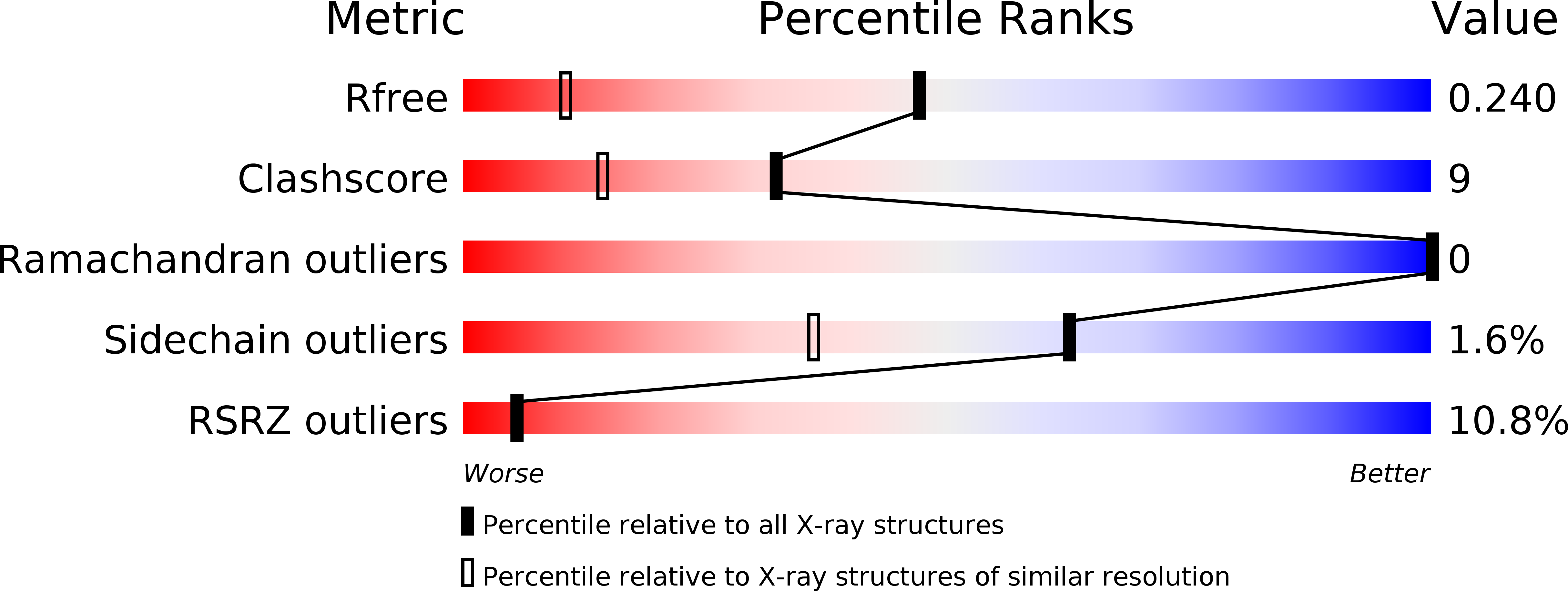
Resolution:
1.50 Å
R-Value Free:
0.22
R-Value Work:
0.20
R-Value Observed:
0.20
Space Group:
P 42 21 2
Macromolecular Entities
Polymer Type:polypeptide(L)
Molecule:TRYPAREDOXIN II
Gene (Uniprot):txnII
Chain IDs:A, B
Chain Length:152
Number of Molecules:2
Biological Source:CRITHIDIA FASCICULATA
Ligand Molecules
Primary Citation
De Novo Phasing of Two Crystal Forms of Tryparedoxin II Using the Anomalous Scattering from S Atoms: A Combination of Small Signal and Medium Resolution Reveals This to be a General Tool for Solving Protein Crystal Structures
Acta Crystallogr.,Sect.D 58 21 ? (2002)
PMID: 11752776 DOI: 10.1107/S0907444901016808

Abstact

The de novo phasing of the structures of two crystal forms of tryparedoxin II from Crithidia fasciculata has been carried out using single-wavelength anomalous diffraction techniques exploiting only the small anomalous signal from the S atoms intrinsic to the native protein. Data were collected at 1.77 A wavelength, where the Bijvoet ratio is approximately 1.2%. Data collected to d(min) = 2.5 A from a crystal of form I, which has a diffraction limit of d(min) = 1.5 A and a solvent content of approximately 46%, produced readily interpretable electron-density maps. When these phases were extended to the resolution limit of the crystals, almost the entire model could be traced automatically. Crystals of form II have a much higher solvent content, approximately 72%, and a much lower diffraction limit than form I and at 1.77 A wavelength yielded data only to d(min) = 2.7 A. Despite the medium resolution of the data for this crystal form, it was possible both to determine the heavy-atom partial structure and then use it to produce, still at d(min) = 2.7 A, an excellent quality interpretable electron-density map. This was then improved by phase extension to the d(min) = 2.35 A diffraction limits of a different crystal for which data were collected on a more intense beamline. The success of this latter structure solution markedly increases the potential use in macromolecular crystal structure determination of the anomalous signal available from S atoms that occur naturally in proteins and, as is discussed, has significant implications for structure determination in the high-throughput era.

Legend

Protein

Chemical

Disease

Primary Citation of related structures