1O7L image
Deposition Date 2002-11-08
Release Date 2003-02-20
Last Version Date 2023-12-13
Entry Detail
PDB ID:
1O7L
Title:
Molybdate-activated form of ModE from Escherichia coli
Biological Source:
Source Organism:
ESCHERICHIA COLI (Taxon ID: 562)
Host Organism:
Method Details:
Experimental Method:
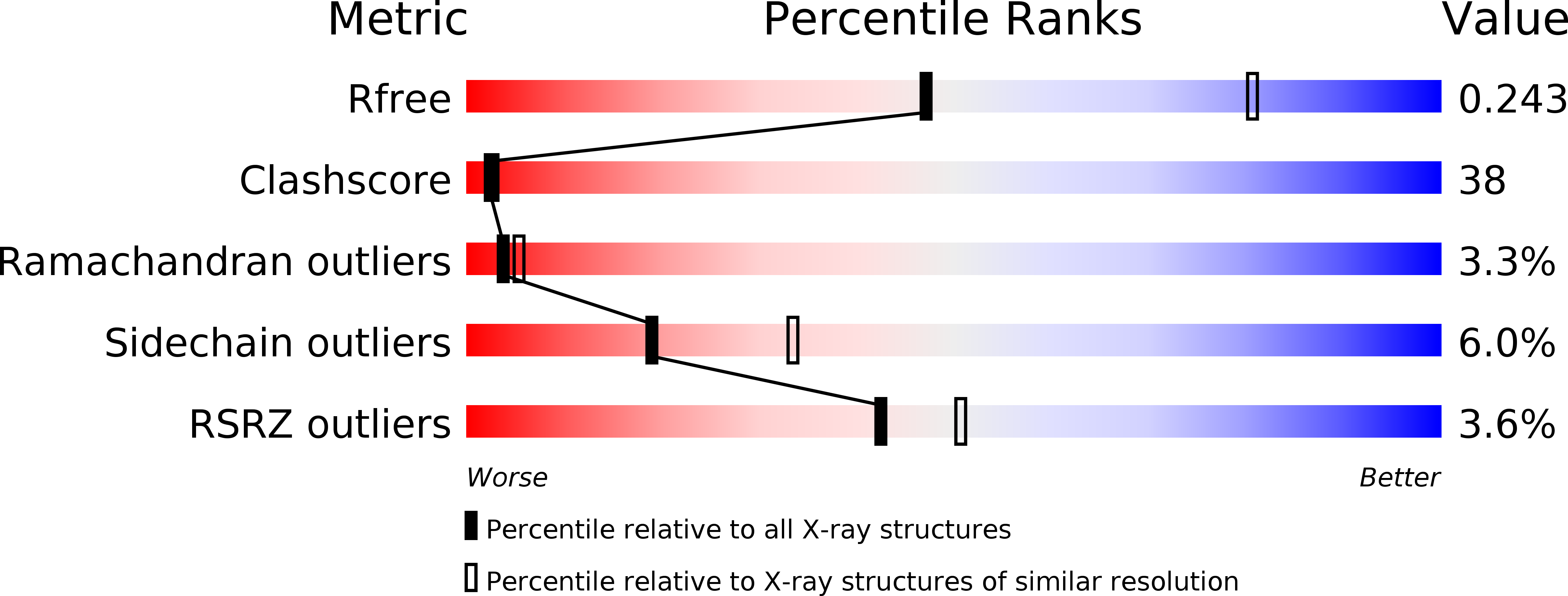
Resolution:
2.75 Å
R-Value Free:
0.25
R-Value Work:
0.20
R-Value Observed:
0.20
Space Group:
P 43
Macromolecular Entities
Structures with similar UniProt ID
Protein Blast
Polymer Type:polypeptide(L)
Molecule:TRANSCRIPTIONAL REGULATOR MODE
Chain IDs:A, B, C, D
Chain Length:262
Number of Molecules:4
Biological Source:ESCHERICHIA COLI
Primary Citation
Crystal Structure of Activated Mode Reveals Conformational Changes Involving Both Oxyanion and DNA-Binding Domains
J.Mol.Biol. 326 761 ? (2003)
PMID: 12581638 DOI: 10.1016/S0022-2836(02)01358-X

Abstact

ModE is a bacterial transcriptional regulator that orchestrates many aspects of molybdenum metabolism by binding to specific DNA sequences in a molybdate-dependent fashion. We present the crystal structure of Escherichia coli ModE in complex with molybdate, which was determined at 2.75A from a merohedrally twinned crystal (twin fraction approximately 0.30) with space group P4(3). We now have structures of ModE in both its "switched on" (ligand-bound) and "switched off" (apo) states. Comparison with the apo structure shows that ligand binding leads to extensive conformational changes not only in the molybdate-binding domain, but also in the DNA-binding domain. The most obvious difference is the loss of the pronounced asymmetry between the two chains of the ModE dimer, which had been a characteristic property of the apo structure. Another major change concerns the relative orientation of the two DNA-interacting winged helix-turn-helix motifs. Manual docking of an idealized DNA structure suggests that this conformational change should improve DNA binding of the activated molybdate-bound ModE.

Legend

Protein

Chemical

Disease

Primary Citation of related structures