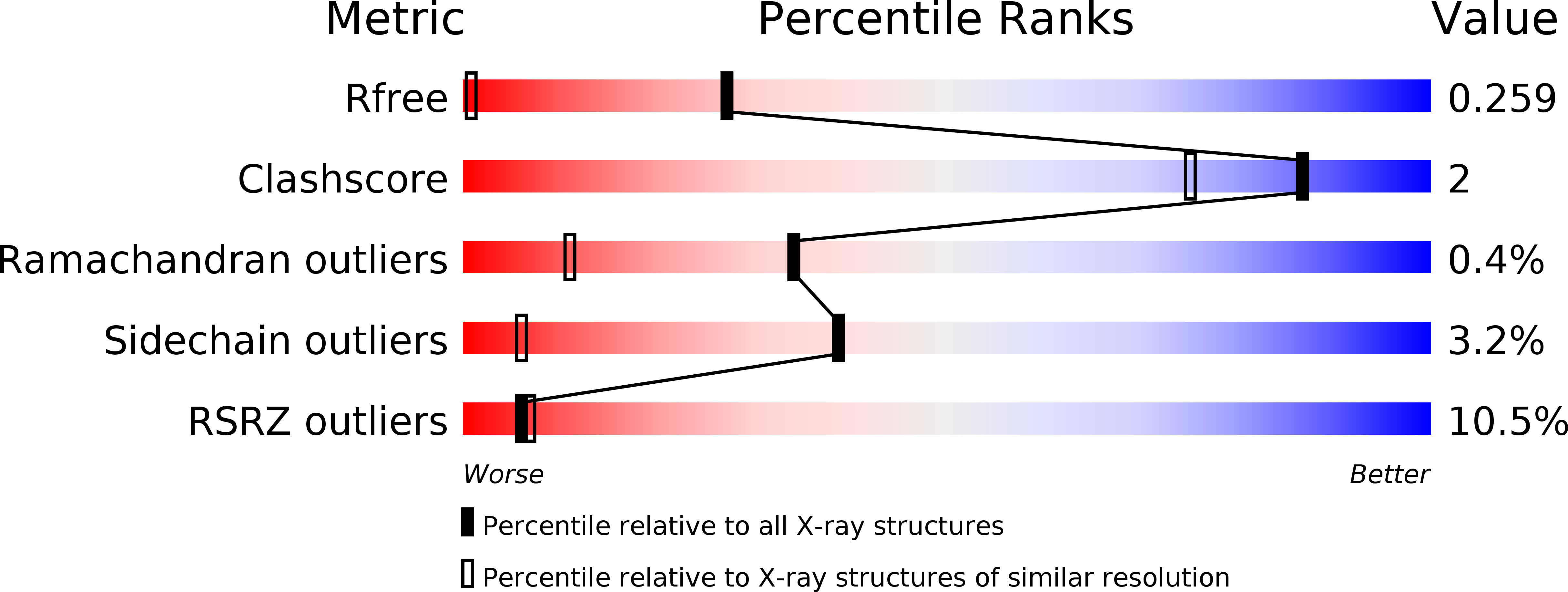
1O7I image
Deposition Date 2002-11-05
Release Date 2003-06-25
Last Version Date 2024-05-08
Entry Detail
PDB ID:
1O7I
Title:
Crystal structure of a single stranded DNA binding protein
Biological Source:
Source Organism:
Host Organism:
Method Details:
Experimental Method:
Resolution:
1.20 Å
R-Value Free:
0.21
R-Value Work:
0.19
R-Value Observed:
0.19
Space Group:
P 61
Macromolecular Entities
Structures with similar UniProt ID
Protein Blast
Polymer Type:polypeptide(L)
Molecule:SINGLE STRANDED DNA BINDING PROTEIN
Gene (Uniprot):ssb
Chain IDs:A, B
Chain Length:119
Number of Molecules:2
Biological Source:SULFOLOBUS SOLFATARICUS
Ligand Molecules
Primary Citation
Insights into ssDNA recognition by the OB fold from a structural and thermodynamic study of Sulfolobus SSB protein.
EMBO J. 22 2561 2570 (2003)
PMID: 12773373 DOI: 10.1093/emboj/cdg272

Abstact

Information processing pathways such as DNA replication are conserved in eukaryotes and archaea and are significantly different from those found in bacteria. Single-stranded DNA-binding (SSB) proteins (or replication protein A, RPA, in eukaryotes) play a central role in many of these pathways. However, whilst euryarchaea have a eukaryotic-type RPA homologue, crenarchaeal SSB proteins appear much more similar to the bacterial proteins, with a single OB fold for DNA binding and a flexible C-terminal tail that is implicated in protein-protein interactions. We have determined the crystal structure of the SSB protein from the crenarchaeote Sulfolobus solfataricus to 1.26 A. The structure shows a striking and unexpected similarity to the DNA-binding domains of human RPA, providing confirmation of the close relationship between archaea and eukaryotes. The high resolution of the structure, together with thermodynamic and mutational studies of DNA binding, allow us to propose a molecular basis for DNA binding and define the features required for eukaryotic and archaeal OB folds.

Legend

Protein

Chemical

Disease

Primary Citation of related structures