1O7D image
Deposition Date 2002-10-30
Release Date 2003-03-20
Last Version Date 2024-10-09
Entry Detail
PDB ID:
1O7D
Keywords:
Title:
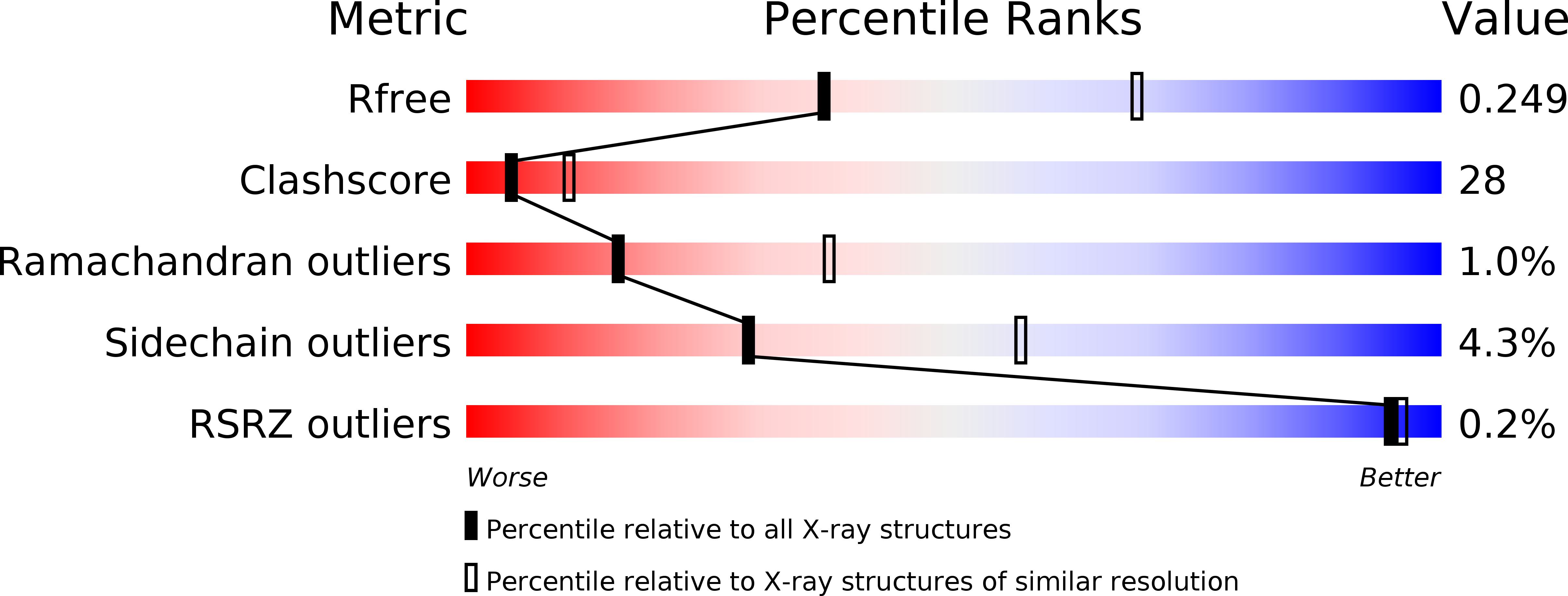
The structure of the bovine lysosomal a-mannosidase suggests a novel mechanism for low pH activation
Biological Source:
Source Organism:
Bos taurus (Taxon ID: 9913)
Method Details:
Experimental Method:
Resolution:
2.70 Å
R-Value Free:
0.28
R-Value Work:
0.25
R-Value Observed:
0.25
Space Group:
P 61 2 2
Macromolecular Entities
Polymer Type:polypeptide(L)
Molecule:Lysosomal alpha-mannosidase
Gene (Uniprot):MAN2B1
Chain IDs:A
Chain Length:298
Number of Molecules:1
Biological Source:Bos taurus
Polymer Type:polypeptide(L)
Molecule:Lysosomal alpha-mannosidase
Gene (Uniprot):MAN2B1
Chain IDs:B
Chain Length:84
Number of Molecules:1
Biological Source:Bos taurus
Polymer Type:polypeptide(L)
Molecule:Lysosomal alpha-mannosidase
Gene (Uniprot):MAN2B1
Chain IDs:C
Chain Length:159
Number of Molecules:1
Biological Source:Bos taurus
Polymer Type:polypeptide(L)
Molecule:Lysosomal alpha-mannosidase
Gene (Uniprot):MAN2B1
Chain IDs:D
Chain Length:282
Number of Molecules:1
Biological Source:Bos taurus
Polymer Type:polypeptide(L)
Molecule:Lysosomal alpha-mannosidase
Gene (Uniprot):MAN2B1
Chain IDs:E
Chain Length:126
Number of Molecules:1
Biological Source:Bos taurus
Modified Residue
Compound ID Chain ID Parent Comp ID Details 2D Image
ASN A ASN GLYCOSYLATION SITE
Primary Citation
The Structure of Bovine Lysosomal Alpha-Mannosidase Suggests a Novel Mechanism for Low-Ph Activation
J.Mol.Biol. 327 631 ? (2003)
PMID: 12634058 DOI: 10.1016/S0022-2836(03)00172-4

Abstact

Lysosomal alpha-mannosidase (LAM: EC 3.2.1.24) belongs to the sequence-based glycoside hydrolase family 38 (GH38). Two other mammalian GH38 members, Golgi alpha-mannosidase II (GIIAM) and cytosolic alpha-mannosidase, are expressed in all tissues. In humans, cattle, cat and guinea pig, lack of lysosomal alpha-mannosidase activity causes the autosomal recessive disease alpha-mannosidosis. Here, we describe the three-dimensional structure of bovine lysosomal alpha-mannosidase (bLAM) at 2.7A resolution and confirm the solution state dimer by electron microscopy. We present the first structure of a mammalian GH38 enzyme that offers indications for the signal areas for mannose phosphorylation, suggests a previously undetected mechanism of low-pH activation and provides a template for further biochemical studies of the family 38 glycoside hydrolases as well as lysosomal transport. Furthermore, it provides a basis for understanding the human form of alpha-mannosidosis at the atomic level. The atomic coordinates and structure factors have been deposited in the Protein Data Bank (accession codes 1o7d and r1o7dsf).

Legend

Protein

Chemical

Disease

Primary Citation of related structures