
Deposition Date
2003-04-20
Release Date
2003-08-26
Last Version Date
2023-12-27
Entry Detail
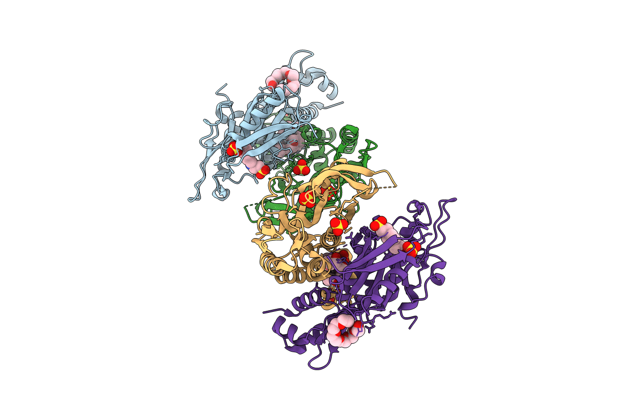
PDB ID:
1O57
Keywords:
Title:
CRYSTAL STRUCTURE OF THE PURINE OPERON REPRESSOR OF BACILLUS SUBTILIS
Biological Source:
Source Organism(s):
Bacillus subtilis (Taxon ID: 1423)
Expression System(s):
Method Details:
Experimental Method:
Resolution:
2.20 Å
R-Value Free:
0.23
R-Value Work:
0.18
R-Value Observed:
0.1
Space Group:
P 1
