1O0R image
Deposition Date 2003-02-24
Release Date 2003-03-04
Last Version Date 2024-10-16
Entry Detail
PDB ID:
1O0R
Keywords:
Title:
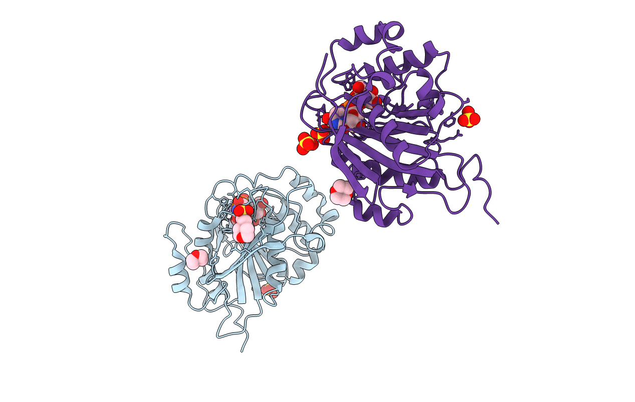
Crystal structure of the catalytic domain of bovine beta1,4-galactosyltransferase complex with UDP-galactose
Biological Source:
Source Organism:
Bos taurus (Taxon ID: 9913)
Host Organism:
Method Details:
Experimental Method:
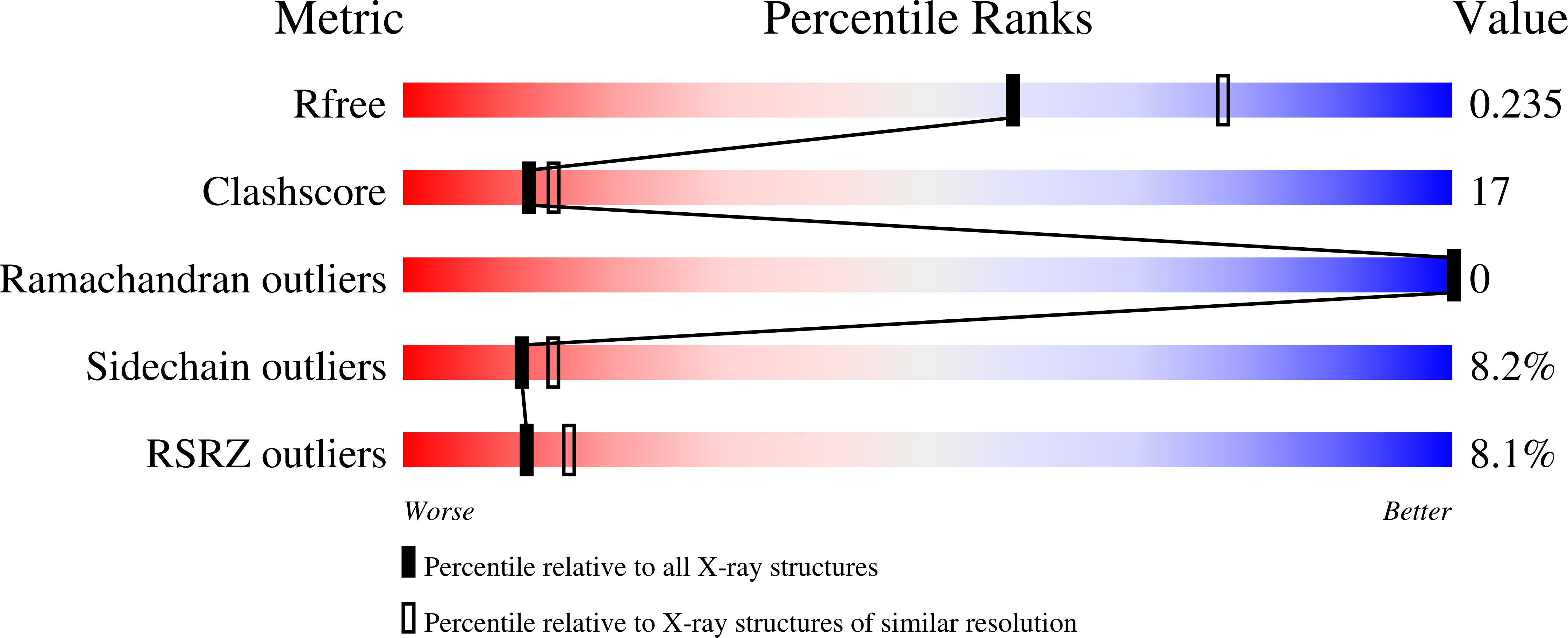
Resolution:
2.30 Å
R-Value Free:
0.25
R-Value Work:
0.20
R-Value Observed:
0.20
Space Group:
P 21 21 21
Macromolecular Entities
Polymer Type:polypeptide(L)
Molecule:beta-1,4-galactosyltransferase
Gene (Uniprot):B4GALT1
Mutations:C342T
Chain IDs:A, B
Chain Length:286
Number of Molecules:2
Biological Source:Bos taurus
Primary Citation
CRYSTAL STRUCTURE OF BETA 1,4-GALACTOSYLTRANSFERASE COMPLEX WITH UDP-GAL REVEALS AN OLIGOSACCHARIDE ACCEPTOR BINDING SITE
J.Mol.Biol. 318 491 502 (2002)
PMID: 12051854 DOI: 10.1016/S0022-2836(02)00020-7

Abstact

The crystal structure of the catalytic domain of bovine beta1,4-galactosyltransferase (Gal-T1) co-crystallized with UDP-Gal and MnCl(2) has been solved at 2.8 A resolution. The structure not only identifies galactose, the donor sugar binding site in Gal-T1, but also reveals an oligosaccharide acceptor binding site. The galactose moiety of UDP-Gal is found deep inside the catalytic pocket, interacting with Asp252, Gly292, Gly315, Glu317 and Asp318 residues. Compared to the native crystal structure reported earlier, the present UDP-Gal bound structure exhibits a large conformational change in residues 345-365 and a change in the side-chain orientation of Trp314. Thus, the binding of UDP-Gal induces a conformational change in Gal-T1, which not only creates the acceptor binding pocket for N-acetylglucosamine (GlcNAc) but also establishes the binding site for an extended sugar acceptor. The presence of a binding site that accommodates an extended sugar offers an explanation for the observation that an oligosaccharide with GlcNAc at the non-reducing end serves as a better acceptor than the monosaccharide, GlcNAc. Modeling studies using oligosaccharide acceptors indicate that a pentasaccharide, such as N-glycans with GlcNAc at their non-reducing ends, fits the site best. A sequence comparison of the human Gal-T family members indicates that although the binding site for the GlcNAc residue is highly conserved, the site that binds the extended sugar exhibits large variations. This is an indication that different Gal-T family members prefer different types of glycan acceptors with GlcNAc at their non-reducing ends.

Legend

Protein

Chemical

Disease

Primary Citation of related structures