
Deposition Date
1994-12-09
Release Date
1995-02-27
Last Version Date
2024-10-16
Entry Detail
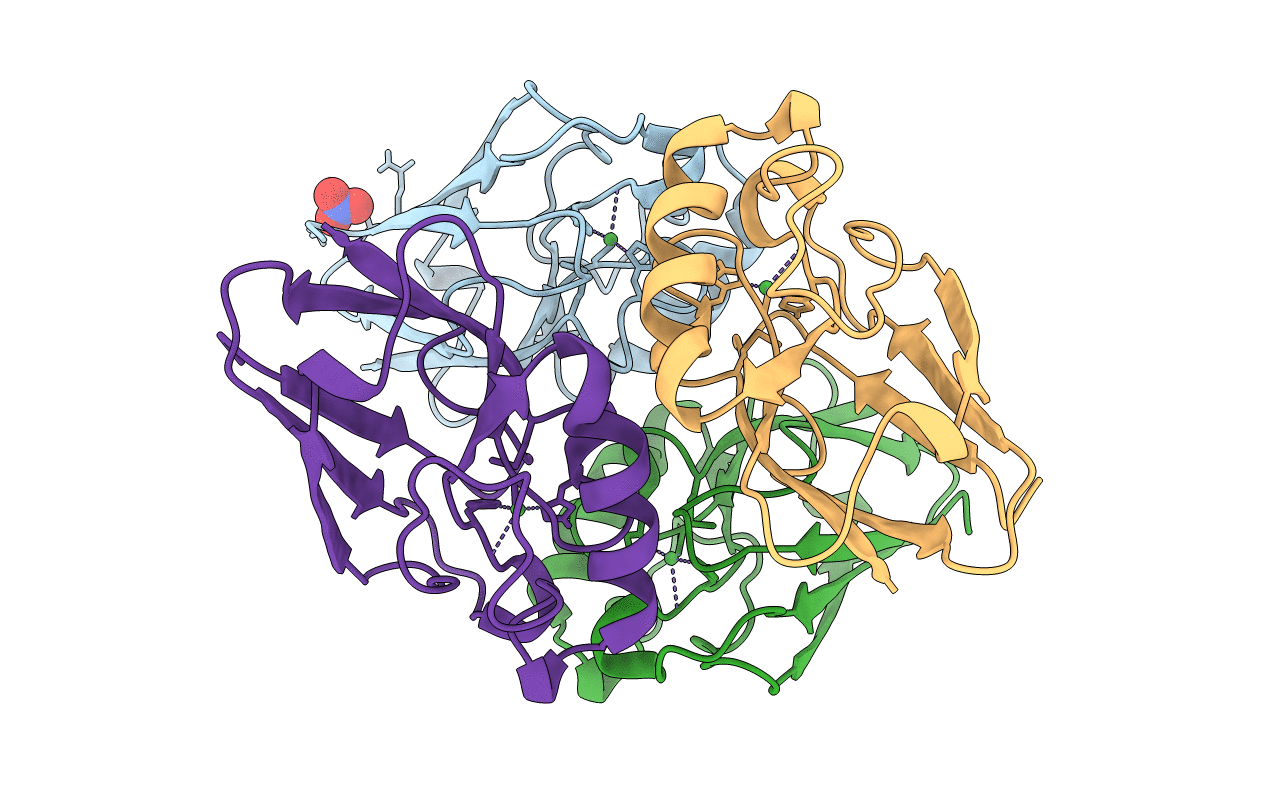
PDB ID:
1NZR
Keywords:
Title:
CRYSTAL STRUCTURE OF THE AZURIN MUTANT NICKEL-TRP48MET FROM PSEUDOMONAS AERUGINOSA AT 2.2 ANGSTROMS RESOLUTION
Biological Source:
Source Organism(s):
Pseudomonas aeruginosa (Taxon ID: 287)
Method Details:
Experimental Method:
Resolution:
2.20 Å
R-Value Work:
0.17
R-Value Observed:
0.17
Space Group:
P 21 21 21
