Deposition Date
2003-02-18
Release Date
2003-04-22
Last Version Date
2024-10-16
Entry Detail
PDB ID:
1NZL
Keywords:
Title:
Crystal Structure of Src SH2 domain bound to doubly phosphorylated peptide PQpYEpYIPI
Biological Source:
Source Organism:
Rous sarcoma virus (strain Schmidt-Ruppin) (Taxon ID: 11889)
Host Organism:
Method Details:
Experimental Method:
Resolution:
1.90 Å
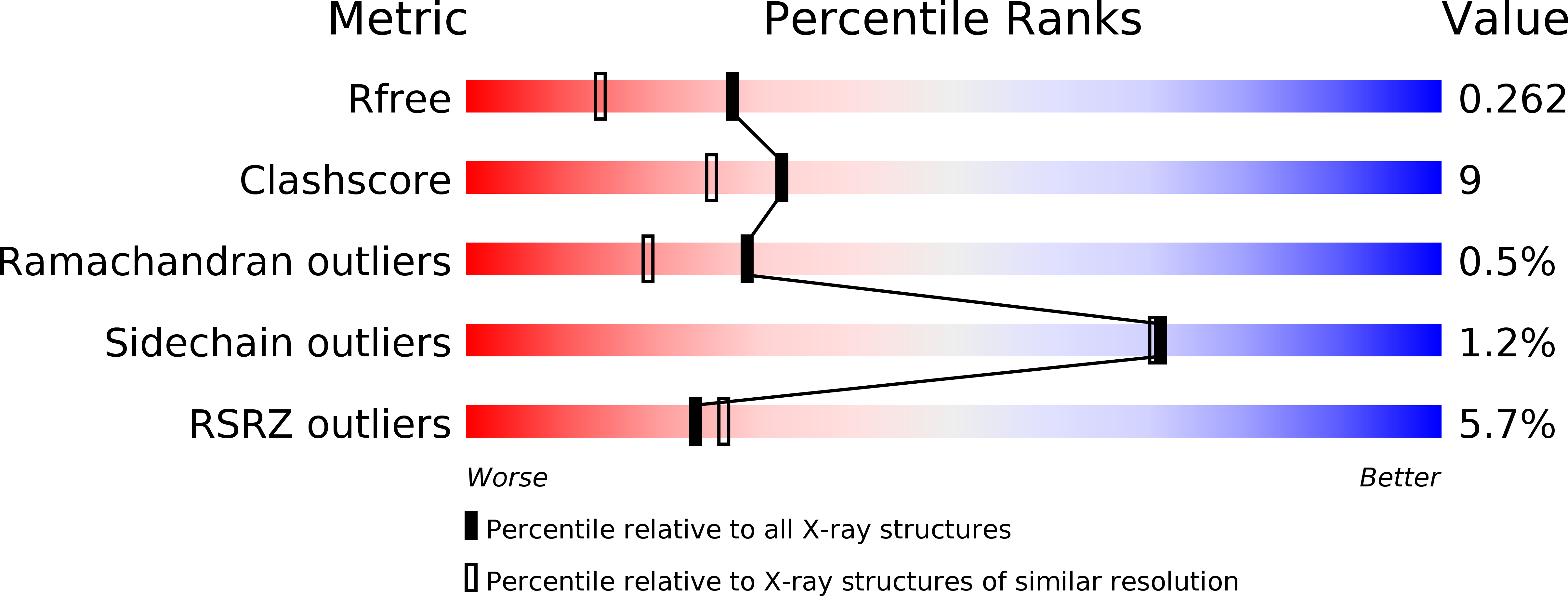
R-Value Free:
0.26
R-Value Work:
0.22
R-Value Observed:
0.22
Space Group:
P 21 21 2