1NZI image
Deposition Date 2003-02-18
Release Date 2003-06-10
Last Version Date 2024-10-23
Entry Detail
PDB ID:
1NZI
Keywords:
Title:
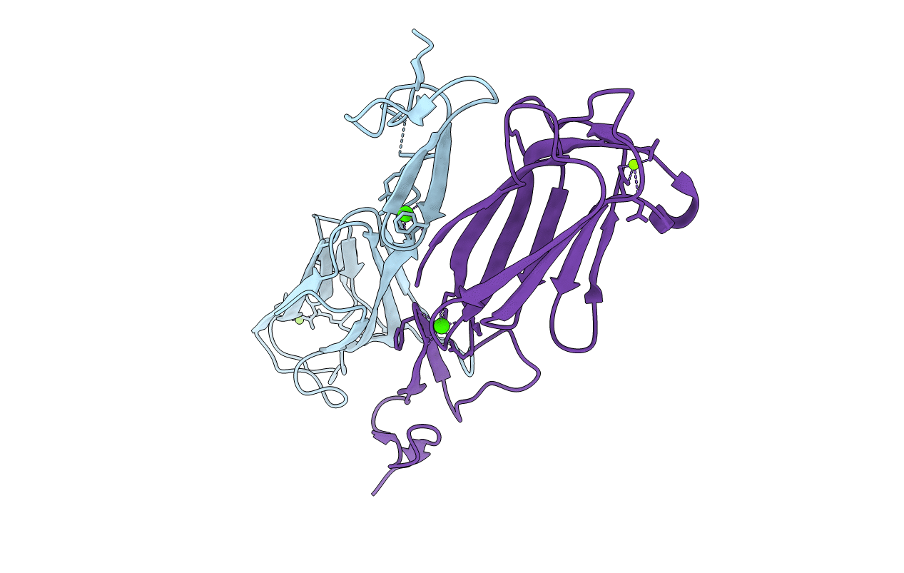
Crystal Structure of the CUB1-EGF Interaction Domain of Complement Protease C1s
Biological Source:
Source Organism:
Homo sapiens (Taxon ID: 9606)
Host Organism:
Method Details:
Experimental Method:
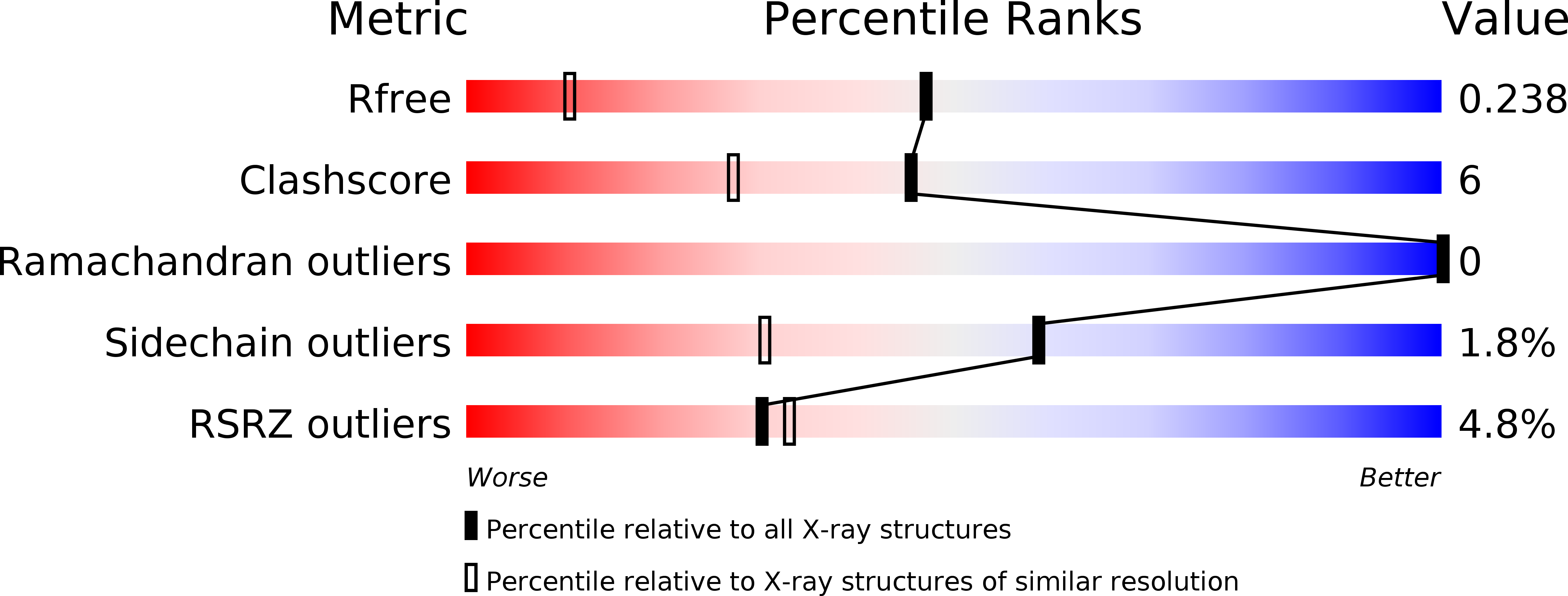
Resolution:
1.50 Å
R-Value Free:
0.23
R-Value Work:
0.21
R-Value Observed:
0.21
Space Group:
P 1
Macromolecular Entities
Structures with similar UniProt ID
Protein Blast
Polymer Type:polypeptide(L)
Molecule:Complement C1s component
Gene (Uniprot):C1S
Chain IDs:A, B
Chain Length:159
Number of Molecules:2
Biological Source:Homo sapiens
Primary Citation
X-ray structure of the Ca2+-binding interaction domain of C1s. Insights into the assembly of the C1 complex of complement
J.Biol.Chem. 278 32157 32164 (2003)
PMID: 12788922 DOI: 10.1074/jbc.M305175200

Abstact

C1, the complex that triggers the classical pathway of complement, is assembled from two modular proteases C1r and C1s and a recognition protein C1q. The N-terminal CUB1-EGF segments of C1r and C1s are key elements of the C1 architecture, because they mediate both Ca2+-dependent C1r-C1s association and interaction with C1q. The crystal structure of the interaction domain of C1s has been solved and refined to 1.5 A resolution. The structure reveals a head-to-tail homodimer involving interactions between the CUB1 module of one monomer and the epidermal growth factor (EGF) module of its counterpart. A Ca2+ ion is bound to each EGF module and stabilizes both the intra- and inter-monomer interfaces. Unexpectedly, a second Ca2+ ion is bound to the distal end of each CUB1 module, through six ligands contributed by Glu45, Asp53, Asp98, and two water molecules. These acidic residues and Tyr17 are conserved in approximately two-thirds of the CUB repertoire and define a novel, Ca2+-binding CUB module subset. The C1s structure was used to build a model of the C1r-C1s CUB1-EGF heterodimer, which in C1 connects C1r to C1s and mediates interaction with C1q. A structural model of the C1q/C1r/C1s interface is proposed, where the rod-like collagen triple helix of C1q is accommodated into a groove along the transversal axis of the C1r-C1s heterodimer.

Legend

Protein

Chemical

Disease

Primary Citation of related structures