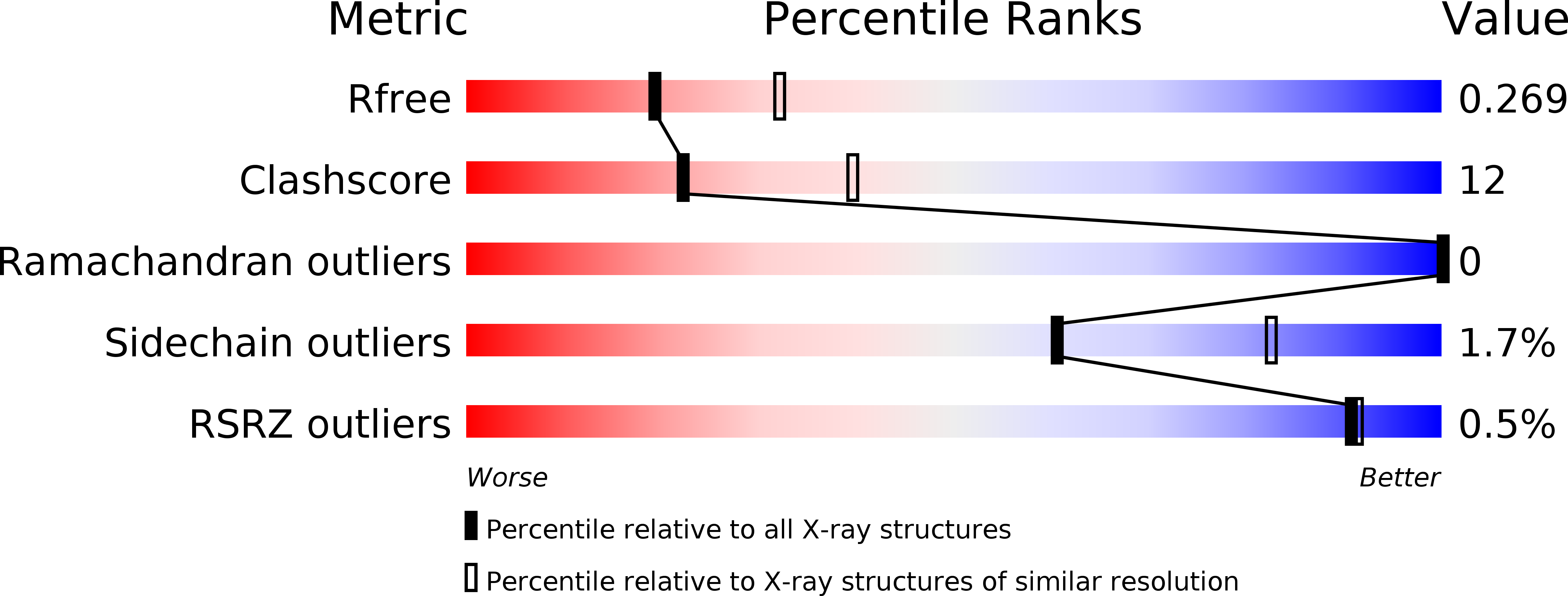
1NZ6 image
Deposition Date 2003-02-16
Release Date 2003-04-22
Last Version Date 2024-10-30
Entry Detail
PDB ID:
1NZ6
Keywords:
Title:
Crystal Structure of Auxilin J-Domain
Biological Source:
Source Organism:
Bos taurus (Taxon ID: 9913)
Host Organism:
Method Details:
Experimental Method:
Resolution:
2.50 Å
R-Value Free:
0.26
R-Value Work:
0.23
R-Value Observed:
0.23
Space Group:
C 2 2 21
Macromolecular Entities
Structures with similar UniProt ID
Protein Blast
Polymer Type:polypeptide(L)
Molecule:Auxilin
Gene (Uniprot):DNAJC6
Chain IDs:A, B
Chain Length:101
Number of Molecules:2
Biological Source:Bos taurus
Modified Residue
Compound ID Chain ID Parent Comp ID Details 2D Image
MSE A MET SELENOMETHIONINE
Primary Citation
Structure-function analysis of the auxilin J-domain reveals an extended Hsc70 interaction interface.
Biochemistry 42 5748 5753 (2003)
PMID: 12741832 DOI: 10.1021/bi034270g

Abstact

J-domains are widespread protein interaction modules involved in recruiting and stimulating the activity of Hsp70 family chaperones. We have determined the crystal structure of the J-domain of auxilin, a protein which is involved in uncoating clathrin-coated vesicles. Comparison to the known structures of J-domains from four other proteins reveals that the auxilin J-domain is the most divergent of all J-domain structures described to date. In addition to the canonical J-domain features described previously, the auxilin J-domain contains an extra N-terminal helix and a long loop inserted between helices I and II. The latter loop extends the positively charged surface which forms the Hsc70 binding site, and is shown by directed mutagenesis and surface plasmon resonance to contain side chains important for binding to Hsc70.

Legend

Protein

Chemical

Disease

Primary Citation of related structures