1NYU image
Deposition Date 2003-02-13
Release Date 2003-04-08
Last Version Date 2024-11-06
Entry Detail
PDB ID:
1NYU
Title:
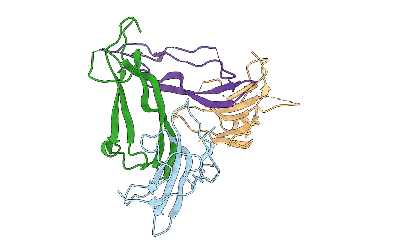
Crystal Structure of Activin A Bound to the ECD of ActRIIB
Biological Source:
Source Organism:
Rattus norvegicus (Taxon ID: 10116)
Homo sapiens (Taxon ID: 9606)
Host Organism:
Method Details:
Experimental Method:
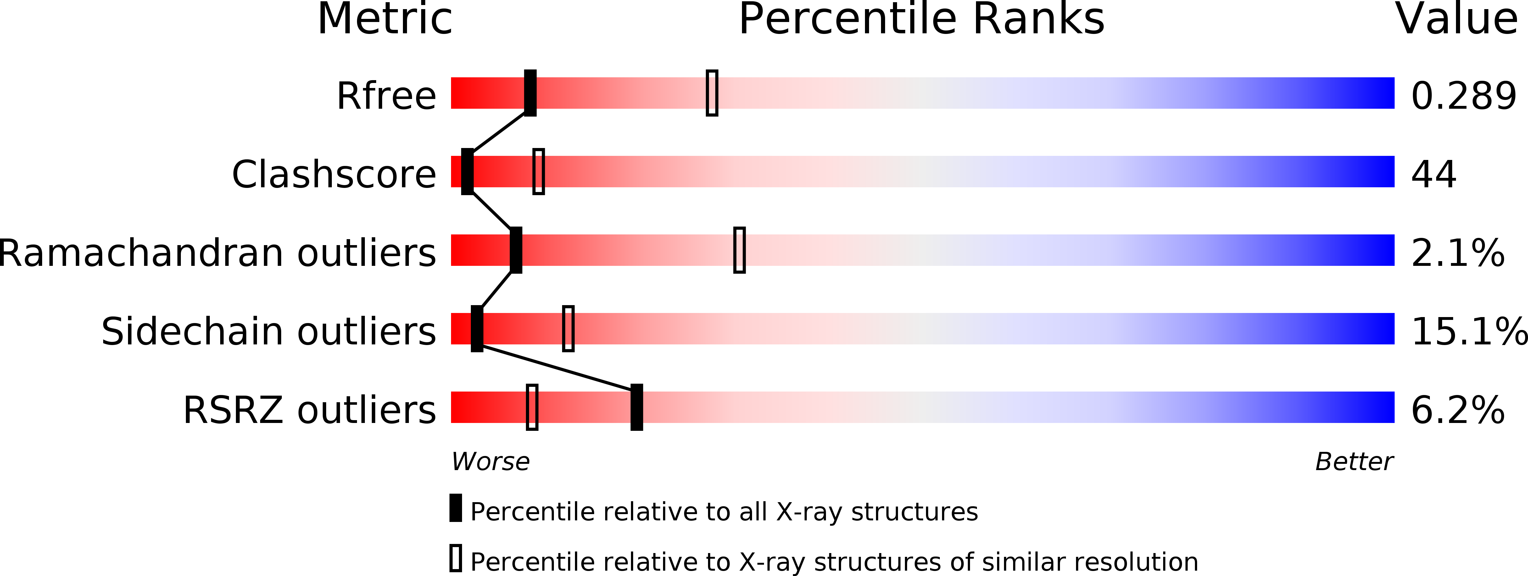
Resolution:
3.10 Å
R-Value Free:
0.29
R-Value Work:
0.26
R-Value Observed:
0.26
Space Group:
P 41 21 2
Macromolecular Entities
Structures with similar UniProt ID
Protein Blast
Polymer Type:polypeptide(L)
Molecule:activin receptor
Gene (Uniprot):Acvr2b
Chain IDs:A, C
Chain Length:105
Number of Molecules:2
Biological Source:Rattus norvegicus
Structures with similar UniProt ID
Protein Blast
Polymer Type:polypeptide(L)
Molecule:Inhibin beta A chain
Gene (Uniprot):INHBA
Chain IDs:B, D
Chain Length:116
Number of Molecules:2
Biological Source:Homo sapiens
Ligand Molecules
Primary Citation
Structures of an ActRIIB:activin A complex reveal a novel binding mode for TGF-beta ligand:receptor interactions
EMBO J. 22 1555 1566 (2003)
PMID: 12660162 DOI: 10.1093/emboj/cdg156

Abstact

The TGF-beta superfamily of ligands and receptors stimulate cellular events in diverse processes ranging from cell fate specification in development to immune suppression. Activins define a major subgroup of TGF-beta ligands that regulate cellular differentiation, proliferation, activation and apoptosis. Activins signal through complexes formed with type I and type II serine/threonine kinase receptors. We have solved the crystal structure of activin A bound to the extracellular domain of a type II receptor, ActRIIB, revealing the details of this interaction. ActRIIB binds to the outer edges of the activin finger regions, with the two receptors juxtaposed in close proximity, in a mode that differs from TGF-beta3 binding to type II receptors. The dimeric activin A structure differs from other known TGF-beta ligand structures, adopting a compact folded-back conformation. The crystal structure of the complex is consistent with recruitment of two type I receptors into a close packed arrangement at the cell surface and suggests that diversity in the conformational arrangements of TGF-beta ligand dimers could influence cellular signaling processes.

Legend

Protein

Chemical

Disease

Primary Citation of related structures
Feedback Form
Name
Email
Institute
Feedback