1NYO image
Deposition Date 2003-02-13
Release Date 2003-08-19
Last Version Date 2024-11-06
Entry Detail
PDB ID:
1NYO
Keywords:
Title:
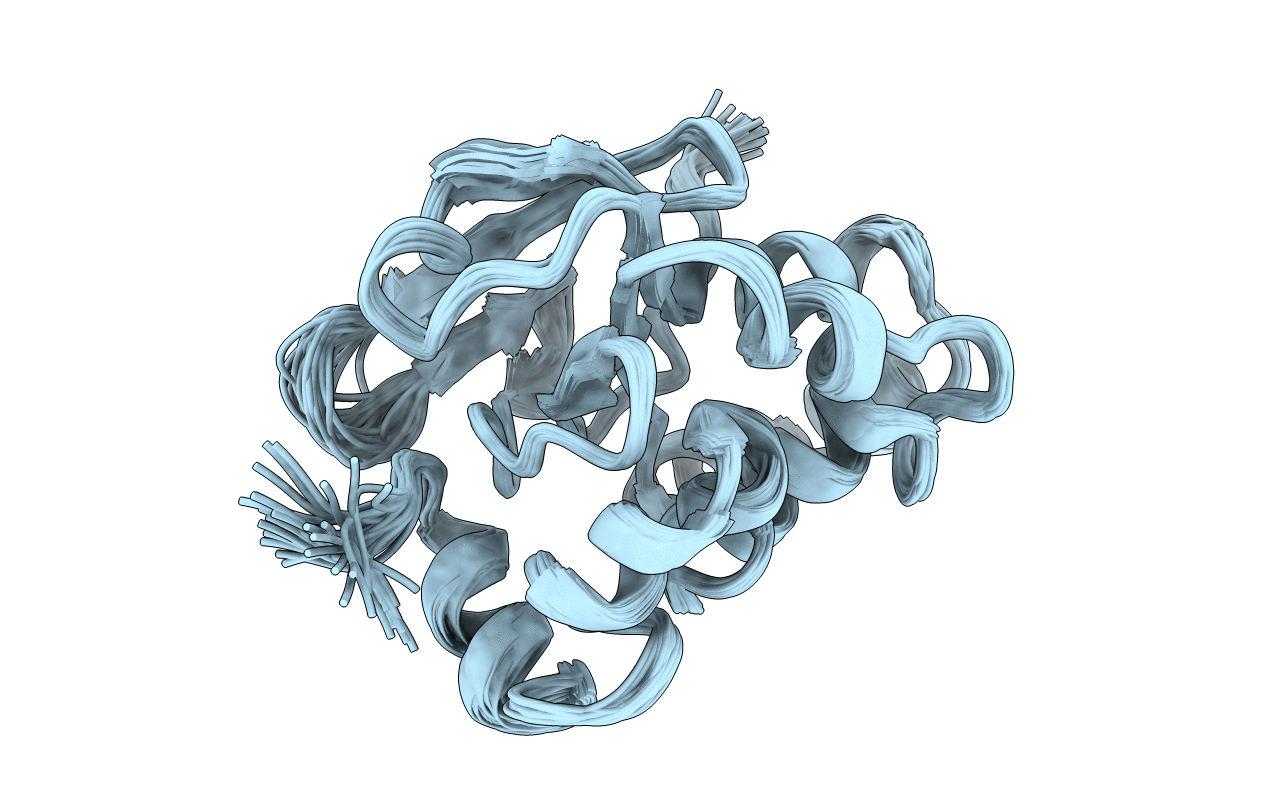
Solution structure of the antigenic TB protein MPT70/MPB70
Biological Source:
Source Organism:
Host Organism:
Method Details:
Experimental Method:
Conformers Calculated:
100
Conformers Submitted:
38
Selection Criteria:
structures with the least restraint violations
Macromolecular Entities
Structures with similar UniProt ID
Protein Blast
Polymer Type:polypeptide(L)
Molecule:Immunogenic protein MPT70
Chain IDs:A
Chain Length:163
Number of Molecules:1
Biological Source:Mycobacterium tuberculosis
Ligand Molecules
Primary Citation
Solution structure of the Mycobacterium tuberculosis complex protein MPB70: from tuberculosis pathogenesis to inherited human corneal desease
J.Biol.Chem. 278 43736 43743 (2003)
PMID: 12917404 DOI: 10.1074/jbc.M307235200

Abstact

The closely related mycobacteria responsible for tuberculosis produce an unusually high number of secreted proteins, many of which are clearly implicated in pathogenesis and protective immunity. Falling within this category are the closely related proteins MPB70 and MPB83. The structure of MPB70 reveals a complex and novel bacterial fold, which has clear structural homology to the two C-terminal FAS1 domains of the cell adhesion protein fasciclin I, whose structures were reported very recently. Assessment of the surface features of MPB70, the sequence divergence between MPB70 and MPB83, the conservation of residues across a group of FAS1 domains, and the locations of disease-inducing mutations in betaig-h3 strongly suggests that MPB70 and MPB83 contain two functional surfaces on opposite faces, which are probably involved in binding to host cell proteins. This analysis also suggests that these functional surfaces are retained in the FAS1 proteins associated with mediating interactions between cells and the extracellular matrix (fasciclin I, periostin, and betaig-h3) and furthermore that some of the human corneal disease-inducing substitutions identified in betaig-h3 will perturb interactions at these sites.

Legend

Protein

Chemical

Disease

Primary Citation of related structures